“Common departments a genetic lab should have are cytogenetics, molecular genetics and molecular cytogenetics. Each department has different testing modules, let’s see what’s inside.”
Genetic and DNA testing has become popular every day. It’s not restricted to genetics; fields like biotechnology, microbiology, embryology, clinical testing, healthcare, evolutionary and population studies also highly rely on genetic testing.
Thus, genetic lab setup should be an integral part of this and many other biological fields. However, setting up a genetic lab isn’t that easy. So we are starting a series named “Genetic Lab Tour” in which we will write articles to show the genetic lab and how it works hypothetically.
This one is the first installment of the series in which I will explain various departments and respective testing facilities for a genetic lab. This will help you to make a decision on which facility you require at your place.
Stay tuned.
Key Topics:
Departments in a Genetic Lab:
Common departments in a genetic lab are cytogenetics, molecular genetics, molecular cytogenetics and others.
Cytogenetic department
Cytogenetics is an interdisciplinary field of genetics, that focuses on the study of chromosomes and related alterations, and their inheritance. It mainly studies chromosomes using staining and banding techniques.
Chromosomes are a complex network of proteins and DNA. They are located in the cell nucleus and comprise genes and other non-coding DNA sequences. It contains telomeres, centromeres and gene-rich arms in their structure.
The condensed form of the chromosome is ‘observably’ visible only during the metaphase. So freshly collected and dividing cells are used for karyotyping and cytogenetic analysis.
Common procedures for cytogenetic investigation are chronologically listed here.
- Cell culture under sterile conditions and using a culture media.
- Cell incubation at 37°C temperature for 48 to 72 hours.
- Cell harvesting to isolate and collect metaphase cultured cells.
- Hypotonic treatment to prepare cells for analysis.
- Staining and banding to observe cells under a microscope.
- Microscopy (high resolution) to investigate chromosomes and study chromosomal abnormalities.
Samples such as whole blood (lymphocytes), amniotic fluid, bone marrow and chorionic villi can be cultured for cytogenetic investigations.
Related article: 10 Common Lab Techniques to Work in a Genetic Lab.
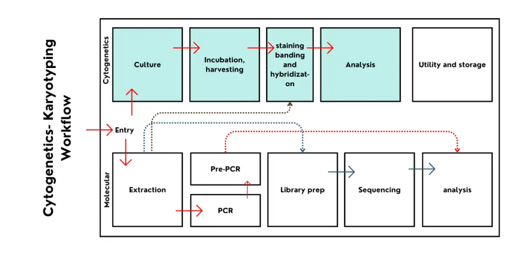
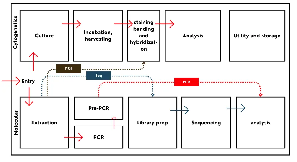
Cytogenetic workflow
Maintaining utmost sterility and contamination-free workflow for a cytogenetic lab is crucial. To do so, a unidirectional workflow should be developed for testing. That means, the sample, at any stage does not flow backward.
Cell culture, incubation, cell harvesting, microscopy preparation and analysis every step should be set up in a separate room to avoid contamination. Cell harvesting, staining, and banding can be combined for smaller, tight-budget laboratories.
Incubated and harvested cells have negligible chances of contamination.
What does it test?
Here is an important question: what can a cytogenetic lab test and detect?
The cytogenetic department in a genetic lab is prepared to test chromosomes. Any numerical or structural chromosomal alterations are directly associated with health-related abnormalities. For instance, trisomy 21 (Down syndrome) is associated with mental retardation and Cri-du-chat is a deletion on chromosome 5 associated with complex health-related issues.
Down syndrome is a numerical chromosomal abnormality while Cri-du-chat is a structural chromosomal abnormality. Both are detected using chromosomal analysis which is popularly known as karyotyping.
The analysis resolution of karyotyping is >5 to 10 Mb. That means, it can effectively investigate abnormalities larger than 5 to 10 Mb. So it can not provide any results related to DNA or gene alterations.
Here is the list of investigations that a cytogenetic lab can perform.
| Abnormality Name | Indication | Karyotype Notation |
| Down Syndrome | Trisomy 21 | 47, XX,+21 or 47, XY,+21 |
| Edwards Syndrome | Trisomy 18 | 47,XX,+18 or 47,XY,+18 |
| Patau Syndrome | Trisomy 13 | 47,XX,+13 or 47,XY,+13 |
| Turner Syndrome | Monosomy X | 45,X |
| Klinefelter Syndrome | Extra X chromosome | 47,XXY |
| Triple X Syndrome | Extra X chromosome | 47,XXX |
| XYY Syndrome | Extra Y chromosome | 47,XYY |
| Cri-du-Chat Syndrome | Deletion of chromosome 5p | 46,XX,del(5p) or 46,XY,del(5p) |
| Wolf-Hirschhorn Syndrome | Deletion of chromosome 4p | 46,XX,del(4p) or 46,XY,del(4p) |
| Philadelphia Chromosome | Translocation t(9;22) | 46,XX,t(9;22)(q34;q11) or 46,XY,t(9;22)(q34;q11) |
| DiGeorge Syndrome | Deletion of 22q11.2 | 46,XX,del(22)(q11.2) or 46,XY,del(22)(q11.2) |
| Williams Syndrome | Deletion of 7q11.23 | 46,XX,del(7)(q11.23) or 46,XY,del(7)(q11.23) |
| Prader-Willi Syndrome | Deletion of 15q11-q13 | 46,XX,del(15)(q11q13) or 46,XY,del(15)(q11q13) |
| Angelman Syndrome | Deletion of 15q11-q13 | 46,XX,del(15)(q11q13) or 46,XY,del(15)(q11q13) |
Molecular Genetics Department:
Molecular genetics is an interdisciplinary field that deals with the study of molecules like DNA and RNA. So it mainly studies DNA, RNA, related alterations and their role in diseases.
Nucleotides (Sugar + Phosphate + Nitrogenous base) create a polynucleotide DNA or RNA chain. DNA is a genetic material for us. Any change in DNA structure or nucleotide order causes serious health-related issues.
Such changes are known as mutations and can be inherited– For instance, thalassemia. PCR, DNA sequencing and restriction digestion are common techniques used for molecular genetic testing.
The order of molecular genetic testing includes these steps.
- Sample collection.
- DNA or RNA extraction
- PCR amplification, sequencing or restriction digestion.
- Analysis- gel or capillary electrophoresis.
- Computational investigations to interpret the results.
Any biological sample can be used for DNA-based analysis. However, only a fresh sample is needed for RNA or gene expression analysis. Blood, buccal swabs, amniotic fluid, chorionic villi, solid tissue and bone are common sample types.
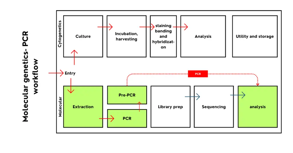
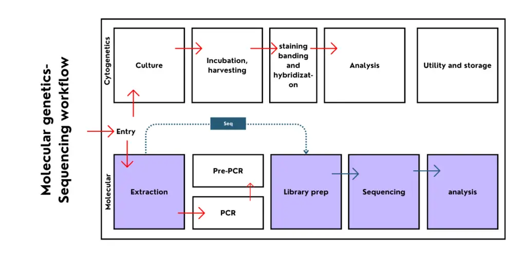
Molecular Genetics Workflow
The molecular genetic workflow is similar to the cytogenetic workflow. It should be in a unidirectional workflow to avoid contamination. Note that the workflow separation varies among testing techniques.
The ideal workflow looks like this– extraction room, pre-PCR room, PCR room, library preparation room, DNA sequencing, post-PCR and sequencing analysis and reporting room.
Related article: Do’s and Don’ts for Genetic Testing Lab Safety.
What does it test?
The molecular genetic department is used to test inherited genetic disorders and health issues in which DNA or gene expression alterations are involved— For example, thalassemia.
It’s an autosomal inherited genetic disorder, caused by mutations in the HBB gene. For analysis, DNA is extracted, HBB gene is amplified using the PCR and sequenced to know the mutations causing the thalassemia.
Common molecular genetic tests are enlisted here.
| Disease Name | Indication | Technique |
| PCR-Based Testing | ||
| Cystic Fibrosis | Mutation in CFTR gene | PCR |
| Huntington’s Disease | Expansion of CAG repeats | PCR |
| Spinal Muscular Atrophy | Deletion of SMN1 gene | PCR |
| Fragile X Syndrome | CGG repeat expansion in FMR1 gene | PCR |
| Sequencing-Based Testing | ||
| Breast Cancer | BRCA1 and BRCA2 mutations | Next-Generation Sequencing (NGS) |
| Lynch Syndrome | Mutations in MLH1, MSH2, MSH6 genes | Sanger Sequencing |
| Marfan Syndrome | Mutations in FBN1 gene | Sanger Sequencing |
| Cardiomyopathy | Mutations in multiple genes (e.g., MYH7, TNNT2) | Next-Generation Sequencing (NGS) |
| Restriction Digestion-Based Testing | ||
| Sickle Cell Anemia | Mutation in HBB gene | Restriction Fragment Length Polymorphism (RFLP) |
| Phenylketonuria (PKU) | Mutations in PAH gene | RFLP |
| Hemophilia A | Mutations in F8 gene | RFLP |
| Tay-Sachs Disease | Mutations in the HEXA gene | RFLP |
Molecular Cytogenetics Department:
Molecular cytogenetics is a department that focuses on cytogenetic investigations at a molecular level. For instance, using fluorescently labeled probes, hybridization can directly be performed on chromosomes.
Such alterations can be studied using a fluorescent microscopy system but can more accurately locate cytogenetic indications than karyotyping. FISH (Fluorescence in situ hybridization) and chromosomal microarray and two common molecular cytogenetic testing scientists use.
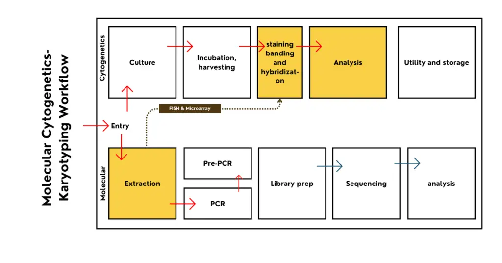
Molecular cytogenetic workflow
The present facility can be combined with a conventional karyotyping setup with several additional workflow additions. For FISH, we need metaphase chromosomes, so for up to metaphase preparation it is combined with karyotyping.
Then a separate room is prepared for hybridization and the fluorescence microscopic analysis can be again combined with the cytogenetics analysis room.
For microarray, the workflow is as stated- DNA extraction, sample preparation and hybridization, microarray array scanning and analysis. Here a few facilities can be combined with the cytogenetics and molecular departments.
Related article: Genomics Lab Setup- Everything You Need to Know To Get Started.
What does it test?
Chromosomal microarray is used to investigate larger indels from all the chromosomes. It can detect various abnormalities associated with different health conditions.
Contrary FISH is used to study various chromosomal alterations. However, for each alteration, a separate FISH probe has been used.
Common molecular cytogenetic tests are enlisted here.
| Disease Name | Indication | Technique |
| FISH (Fluorescence In Situ Hybridization) | ||
| Chronic Myelogenous Leukemia (CML) | BCR-ABL translocation (t(9;22)) | FISH |
| DiGeorge Syndrome | 22q11.2 deletion | FISH |
| Prader-Willi Syndrome | 15q11-q13 deletion | FISH |
| Williams Syndrome | 7q11.23 deletion | FISH |
| Chromosome Microarray | ||
| Autism Spectrum Disorders | Copy number variations (CNVs) | Chromosome Microarray (aCGH) |
| Congenital Heart Defects | Microdeletions and duplications | Chromosome Microarray (aCGH) |
| Epilepsy | Genomic imbalances | Chromosome Microarray (aCGH) |
| Developmental Delays | Microdeletions and duplications | Chromosome Microarray (aCGH) |
Wrapping up:
These are the three common departments any research or diagnosis genetic lab has. This article is prepared for students and professionals to understand the basic structure of a lab.
I hope this article helps you. In the next article, we will discuss the instrumentation for each department.
P. S. Want to set up a genetic lab or upgrade your existing one? Contact our team now!