“Learn how the triple helix DNA formed and the mechanism behind it. Explore the biological significance of triplex helix DNA”
DNA (Deoxyribose nucleic acid) is a fundamental molecule of life and is present in every organism. We know DNA as a storage unit that can also express and transmit genetic information. It plays a major role in biological development and reproduction.
The basic structure of a DNA is simple and consists of only sugar, phosphate and nitrogenous bases. The base pairing between purine and pyrimidine gives DNA a classic double helix structure.
The double-stranded DNA helix is the most stable and common structure present in all organisms. However, it may open up for replication, transcription and translation. The dsDNA structure gives our DNA stability, functionally and enables it to fit in the cell nucleus.
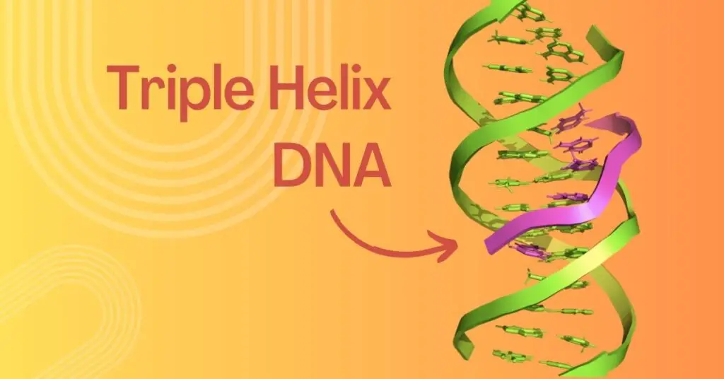
But do you know? Another form of DNA- the triplex helix DNA or three-stranded DNA structure also exists in nature. Triple helix DNA forms when a single-stranded DNA binds with the double-stranded DNA.
So what a triplex helix DNA is, how does it form and what is its biological significance? Let’s find out. In this article, I will explain an unusual DNA structure– the triple helix DNA, its structure, formation and importance.
Stay tuned.
Key Topics:
What is Triple Helix DNA?
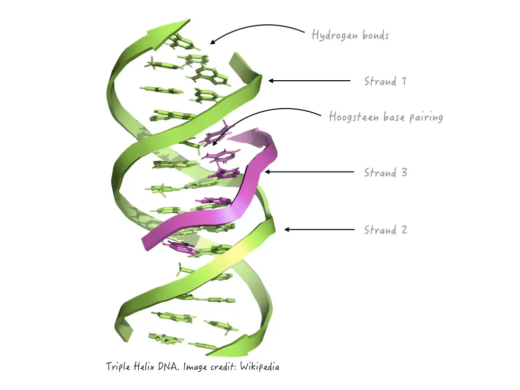
The triple helix is an uncommon, unconventional and atypical DNA form that occurs only when a single strand of DNA binds with the double-stranded DNA by an unusual Hoogsteen hydrogen bonding. Notedly, the third single helix binds largely to DNA major groove.
The idea about the triple helix nucleic acid was given by Linus Pauling in around 1950 when the structure of DNA was being proposed by Watson and Crick. Pauling’s simple demonstration showed that the dsDNA can bind with another single-stranded DNA and form a triple helical structure.
However, the structure was actually (experimentally) demonstrated by Felsenfeld and Rich in 1957. In 1987, Peter Dervan proposed an in vitro model of triple helical DNA using synthetic oligonucleotides which was named TFO.
Triplex-forming nucleotides TFO, which has been used in constructing the triple helix DNA, opened a new door for biotechnology and genetic engineering research. It is used in gene targeting and gene suppression studies and many more.
Now let’s check out how this interaction occurs and how the triple helix DNA forms:
Formation of Triple helix DNA:
The classic double helix DNA follows a Watsons and Crick base pairing rule in which the nitrogenous bases are attached by hydrogen bonds; three between Guanine and Cytosine and two between Adenine and Thymine, respectively and stabilize the dsDNA.
However, any triple helix DNA undergoes Hoogsteen base pairing to form a stable triplex DNA structure. Now let’s first understand the concept of Hoogsteen base pairing so that the mechanism of H-DNA formation can be understood properly.
Hoogsteen base pairing also consists of hydrogen bonding, however, it is entirely different from the conventional Watson and Crick base pairing. Watson and Crick’s pairing typically occurs between Adenine and thymine, and Guanine and Cytosine, and vice versa which gives the DNA a helical structure.
In the Hoogsteen base pairing, the pairing occurs between the purines that face the major groove and the Watson and Crick base pairing of the double-stranded DNA.
Or the purine bases rotate 180° around its glycosyl bond and consequently alter the hydrogen bonding pattern (anti-Hoogsteen base pairing). So the hydrogen bonding pattern changes and occurs at different positions compared to the typical WC base pairing.
The Hoogsten base pairing is an atypical, non-canonical and unconventional type of pairing, and only occurs in altered physiological conditions. It is commonly reported in purine bases- Adenine and Guanine.
So resultantly, Adenine pairs with Cytosine or Thymine while Guanine can pair with Cytosine or Adenine. Such kind of unusual base pairing (Hoogsteen base pairing) is reported when the third single strand will bind with the dsDNA and form the triple helix structure.
The concept was explained by Hoogsteen in 1963. Notedly, Hoogsteen base pairing is less stable and weak which makes the triple helix structure less stable. Although, the stability of the triple helix DNA depends on the length of the triplex formation, and physiological conditions such as pH, salt concentration or temperature of the environment.
This is a simple explanation of this concept. We will cover the entire and comprehensive explanation in some other article.
So the triple helix DNA forms when the purine bases of the third strand (artificially prepared oligonucleotide strand or the strand of the same DNA) form hydrogen bonds (Hoogsteen base pairing) with the purine bases of Watson and Crick base pairing, common at the major DNA groove.
Interesting article: How to Write a DNA Sequence and its Complementary Pairing?
Types of triple helix DNA:
The triplication of DNA can be classified depending on the DNA molecule involved or the type of base pairing present. Intermolecular and intramolecular are two common types of triple helix DNA depending on the DNA molecule involved.
Intramolecular triple helix:
This type of triplex involves a single polynucleotide chain having three nearly complementary regions. The three nearly or completely complementary regions of a long single DNA aligned with each other and forms a triple helix structure.
Or one of the DNA duplexes provides a complementary strand to form the triple-stranded DNA. It is popularly known as H-DNA.
No external single-stranded oligonucleotide is needed to prepare an intramolecular triple helix and thus it can naturally occur in a cell. A single DNA folds back and constructs a triplex structure.
Intermolecular triple helix:
This type of triple helix is formed when a third complementary single DNA strand binds with the duplex DNA using either Hoogsteen or Reverse Hoogsteen base pairing. An artificially synthesized single-stranded DNA, known as triplex-forming oligonucleotide (TFO), complementary to any of the duplex DNA strands binds and forms the triple helix.
The third strand though is parallel to our dsDNA bases, doesn’t follow classic Watson and Crick base pairing, as aforementioned. The concept of Intermolecular triple helix has been popularly utilized in gene therapy, gene targeting and other biotechnology techniques.
Now depending upon the type of base pairing different types of triple DNA helix structures are explained here:
Hoogsteen triple helix:
This is the most common type of triple helix formation and scientists also use it for in vitro experiments. Here as we said, the purine bases of the third strand bind with the purine bases of any of the dsDNA at the DNA major groove.
Reverse Hoogsteen triple helix:
Reverse Hoogsteen base pairing also results in a triplex of strands in which the base pairing occurs when the nucleobases are in reverse orientation. The third strand here mostly binds with the DNA minor groove.
Hoogsteen and Watson and Crick triple helix:
In this type of triple helix, both Hoogsteen and Watson and Crick base pairing are observed. The third strand will bind at two different strands with two different types of base pairing viz. Hoogsteen base pairing with one strand and normal base pairing with another strand.
Quadruplex triple helix:
This type of helix is formed when the third DNA strand binds with the Quadruplex DNA (four-stranded DNA structure). This typically occurs by the association of four different Guanine-rich strands, Hoogsteen base pairing and Watson and Crick base pairing.
Read more: Deciphering The Unique Characteristics of DNA Major and Minor Grooves.
Triple Helix Model:

Biological significance:
Triple helix DNA is an abnormal DNA structure and doesn’t appear normally in our genome. We can consider it as an abnormality in primary DNA structure. It is only reported, however, in a very small genomic portion and under suboptimal conditions.
In humans and other mammals, such a structure is reported, however, is less stable and biologically irrelevant. Interestingly, some organisms may contain a prominent amount of such structures.
For example, the genome of Ciliate Stylonychia Lemnae contains a significant amount of triple helical structures at the telomeric repeat sequences. But, it has not been identified or featured as a natural feature of any organism’s genome.
A high abundance of sequences is capable of forming the H-DNA present in the eukaryotic genome. A study suggests that ~1 in every 50,000 bp sequences is capable enough to form the H-DNA in our genome. However, the major polypurine: polypyrimidine regions are located on non-coding sequences.
Like the Ciliate Stylonychia Lemnae, other organisms including the human genome do have a higher capacity to form the triple helix DNA structure frequently. The biological significance of H-DNA is still not clear. Studies, though, suggest that such structures may form in sub-optimal conditions and can help in DNA repair and gene expression regulation.
Wrapping up:
This is just the beginning, a lot to come on this topic. Triple helix structure is not a natural DNA structure entity, however, scientists use this unusual phenomenon for our benefit. Artificially synthesized oligos can be used as a third strand and used for gene silencing, gene transfer and other applications.
It’s a novel and futuristic tool for various biotechnology, nanotechnology and genetic engineering applications. In the next article on this topic, we will comprehensively take a call on various applications of triple helix DNA structure.
Till then, subscribe to our blog, share this post and do bookmark our blog for more content.


