“Discover the fascinating world of DNA- genetic material that makes us who we are. Learn about the definition, structure and function of DNA and how it determines various traits.”
Do you ever wonder, what makes you, ‘you?’ What makes you unique and different from other individuals or organisms? What defines your eye color, height, skin color, or competency for adaptation and susceptibility/resistance to any disease?
Our DNA, right? A non-scientific person even knows the answer that DNA makes us unique in all senses. It is everything for not only us but every life on earth; hence, every organism carries DNA in its cells. (Note that retrovirus doesn’t have DNA).
DNA doesn’t just make us unique but it is also crucial for us. A bio-molecule– DNA has been present since the very beginning of life on Earth and shaped evolution through various means.
We know DNA as our genetic material and blueprint of life. But do you know what exactly the DNA is? Why is it important? And why is it the genetic material for us? In this article, we will answer all these questions and define the structure, chemical constituents, function and importance of DNA.
Starting from the basics, everything will be covered and explained thoroughly. We’ll also look at the experiments that established its identity as “genetic material” and made it so crucial. I will also list and explain various types of DNA and their importance.
So if you are curious about knowing DNA, well! Stick around. We are going to deep dive into the fascinating world of ‘DNA’, and I promise, you won’t want to miss it! Take a cup of coffee and stay tuned.
Related article: How to Write a DNA Sequence and its Complementary Pairing?
Key Topics:
What is DNA?
DNA is a biomolecule present in the cell and established its identity as our genetic material. Put simply, it’s like a computer hard disk– it stores information for an organism and for every life on Earth and expresses it to survive.
Because it is present in the cell nucleus and acidic in nature, it’s popularly known as nucleic acid. We can say it’s one type of nucleic acid. And thus, the name deoxyribose nucleic acid is given. In 1869, Friedrich Miescher isolated nucleic acid from a cell and named it “nuclei” However, Richard Altmann, who was a student of Miescher F, gave the original name “nucleic acid” in 1888.
Two types of well-established nucleic acids are DNA and RNA. As aforesaid, RNA- as genetic material is only present in retroviruses. DNA is the most prevalent form of genetic material on Earth. But before comprehensively explaining its structure, let’s move through some important historical milestones in the discovery of DNA.
| Year | Name of Scientist(s) | Discovery |
| 1869 | Friedrich Miescher | Discovered ‘nuclein’, later named “nucleic acid” |
| 1888 | Richard Altmann | Characterized nucleic acid. |
| 1910 | Thomas Hunt Morgan | Demonstrated that genes are located on chromosomes. |
| 1913 | Hershey A and Chase M | DNA is a genetic material, not a protein. |
| 1927 | Koltsov N | Chromosomes are made up of DNA. |
| 1928 | Frederick Griffith | Demonstrated that genetic information can be transferred between cells. |
| 1931 | Morgan TH et al. | Genes are arranged linearly on chromosomes. |
| 1944 | Avery O, MacLeod C and McCarty M | DNA is a genetic material and is responsible for the transformation of genetic information between cells. |
| 1951 | Chargaff E | Postulated that amounts of A, T, G and C are not equal. The no of A and T, and G and C are equal. |
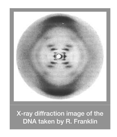
| 1952 | Rosalind Franklin and Wilkins | Captured the image of DNA using X-ray crystallography. |
| 1953 | Watson J and Crick F | Postulated the chemical structure of DNA and decoded the double-helical nature of DNA. |
| 1959 | Arther Korneberg | Discovered DNA polymerase. |
| 1966 | Nirenberg M, Khorana HG and Holley R | Decipher the genetic code. |
These discoveries postulate many facts regarding DNA and falsify many too. Here are some properties of DNA according to these findings.
Properties of DNA:
- DNA is present in the cell nucleus.
- It is known as a nucleic acid which is acidic in nature.
- It stores and transfers genetic information from one to another cell.
- It is located on chromosomes.
- It’s helical in shape and located on chromosomes in a linear fashion.
- The number of adenines is equal to the number of thymine while the number of cytosines is equal to the number of guanines in the DNA.
Now moving ahead, nucleic acid- our DNA is a long chain of nucleotides and hence known as the polynucleotide chain. The concept of nucleotides becomes clear after we discuss the structure. So let’s move on to the shape and size, and structure of DNA.
Shape and Size:
The three major structural constituents of DNA are sugar, phosphate and nitrogenous bases. Bases in DNA create a spiral shape which gives DNA its classic double-helix shape. This structure highly resembles a twisted rope or ladder.
Two individual single-strands form complementary base pairing and create a double-stranded DNA structure, while the uneven arrangement of bases forms major and minor DNA grooves. Both grooves have a pivotal role in the regulation of gene expression.
B-form DNA (we will discuss the types of DNA later in this article), is the most prevalent form in nature. One complete turn is 34Å in length and consists of one major and minor groove. The length between adjacent bases is 3.4Å.
One complete turn consists of roughly ~10-12 bases. The width of the DNA helix is 20Å. Notedly, the size of major and minor grooves also vary. Structurally, the base pairing covers the inner space of the DNA while the phosphate faces outside of the DNA.
Related article: DNA vs RNA: Differences and similarities.
Structure of DNA:
As aforesaid, deoxy sugar, nitrogenous bases and triphosphate are the structural constituents of DNA.
Sugar:
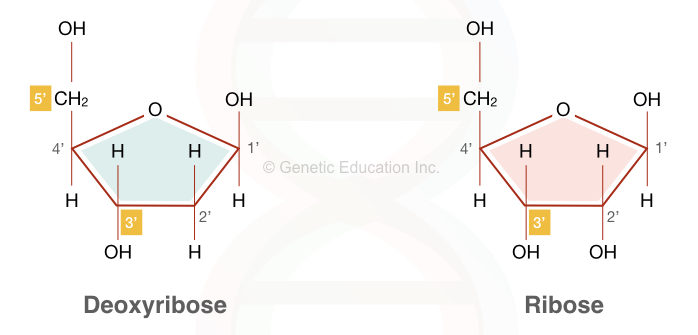
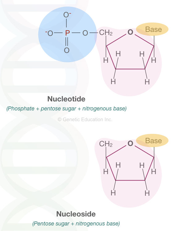
Sugar in nucleic acid is pentose sugar having five carbons. Both DNA and RNA contain the same pentose sugar, however, DNA contains deoxyribose sugar whereas RNA contains only ribose sugar.
The chemical formula of the DNA sugar is 2’ -Deoxy D-ribose while the chemical formula of the RNA sugar is D-ribose. The only difference between DNA and RNA is that the DNA lacks one oxygen at the second carbon position of the pentose ring.
The ribose present in DNA is in beta-furanose forms and arranged in a cyclic manner. Importantly, sugar is the unit that distinguishes DNA from RNA not bases. The 1st and 5th carbons of the sugar are attached with a nitrogenous base and phosphate, respectively.
The structure of the beta-furanose is given below,
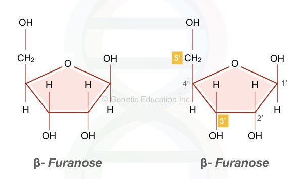
This structure (Sugar + phosphate + nitrogenous base) is collectively known as ‘nucleotide.’ When it lacks phosphate, it is known as ‘nucleoside.’ The structure of deoxyribose and ribose sugar is explained here in the image.
Phosphate:
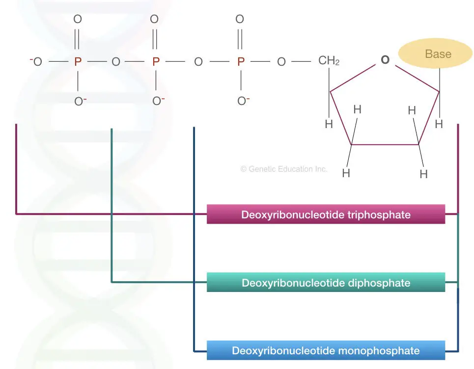
Phosphate is the backbone of DNA and is attached to the deoxyribose at the 5th carbon position. The negative charge of the phosphate makes the DNA net negatively charged molecule. In addition, it’s the phosphate that gives DNA an acidic nature.
Our DNA is deoxyribose nucleotide triphosphate, meaning, three consecutive phosphates are present in the single unit of DNA. If only one or two phosphates are present, they can be known as deoxyribose nucleotide monophosphate and deoxyribose nucleotide diphosphate, respectively.
The structure of mono, di and triphosphate is explained in the image below.
Nitrogenous bases:
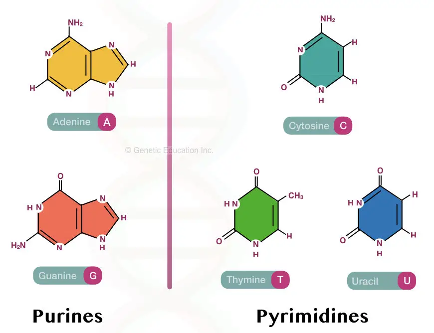
All four nitrogenous bases– A’s, T’s, C’s and G’s are classified into two broad categories– purines and pyrimidines. Adenine and Guanine are purine bases while Cytosine, Thymine and Uracil are pyrimidine bases.
In general, both purines and pyrimidines are heterocyclic and aromatic compounds consisting of rings. Purines have one hexose and one pentose ring while pyrimidines only have a six-carbon hexose ring.
Both contain different numbers of nitrogens in their structure. Purines and Pyrimidines, although, are hydrophobic in nature, can soluble in water at acidic pH. They remain water-insoluble at neutral pH.
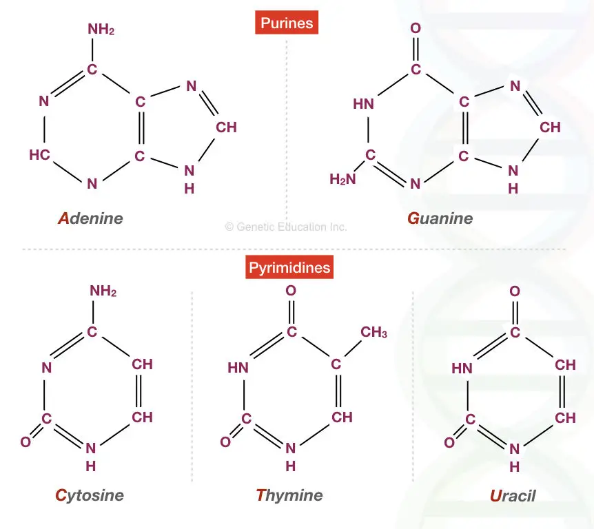
| Purine | Chemical name |
| Guanine | 2-amino-6-oxy purine |
| Adenine | 6-amino purine |
| Pyrimidine | Chemical name |
| Thymine | 2, 4-deoxy-5-methyl pyrimidine |
| Uracil | 2, 4,-deoxy pyrimidine |
| Cytokine | 2-oxy-4-aminopyrimidine |
Want more colorful images? See this image;
Do you know?
The name Thymine is given because it was first isolated from thymus tissues while the Guanine name comes from the Guano bird manure.
Other nitrogenous bases Xanthine and Hypoxanthine –purines and Orotic acid –pyrimidine are also present in nature but not in DNA or RNA.
Now let’s see how bases are attached and form the classic helical structure of the DNA.
Bonds in DNA:
A weak hydrogen bond occurs between nitrogenous bases of the opposite (sense and antisense) strands. Two hydrogen bonds between Adenine and Thymine and three hydrogen bonds between Cytosine and Guanine are present.
Hence, the bonding between C’s and G’s is stronger because of the three hydrogen bonds, however, hydrogen bonding is very weak, comparatively and occurred temporarily. It can easily beak by heating or chemical treatment.
So hydrogen bonding is horizontal and occurs between the bases of sense and antisense strands. It gives the DNA its classic dsDNA helical structure.
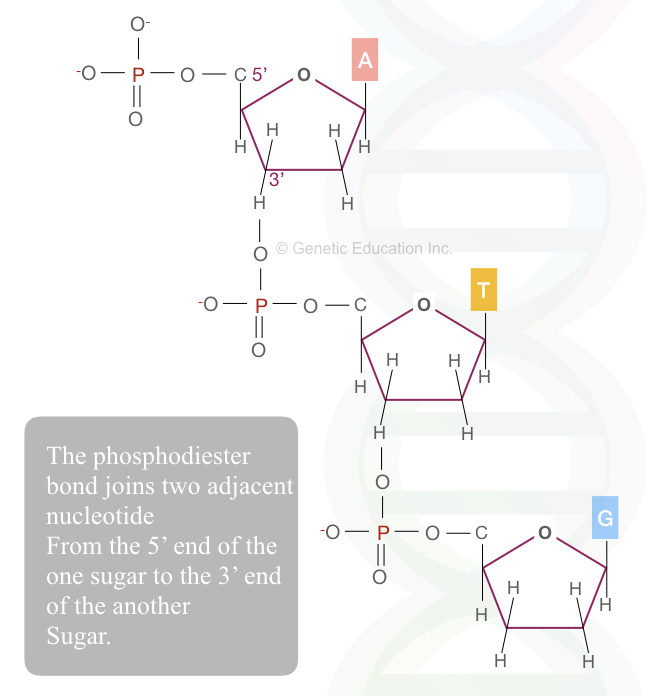
Another important bond in the DNA structure is the phosphodiester bond. It occurs between the 3’ carbon of the one sugar and the 5’ carbon of the second sugar in the same plane. It’s a vertical bonding between the sugar of the same strand. So all the phosphodiester bonds are in the same orientation along the chain.
Besides these two crucial bonds, another bond named, the “glycosidic bond” is present between the pentose sugar and nitrogenous bases. Collectively, all the bonds together form a stable, more electronegative and double-helical DNA structure.
Studies also suggest that weak Van der Waal forces, base-stacking interactions and a weak hydrophobic effect between DNA bases also help stabilize the duplex DNA.
Definition:
“DNA is a nucleic acid made up of sugar, phosphate and nitrogenous bases that stores, express and inherits genetic information.”
Functions of DNA:
Three primitive functions of DNA are to store, express and transfer genetic information from one to the next generation. This allows us to survive, express traits, produce new traits, create diversity, reproduce and sustain the continuity of life on Earth.
So life can only be sustained and survived by DNA and thus, it’s known as life’s blueprint. Now take a look at how these functions are executed.
Encode genetic information:
Genetic information is our source of ‘commands’ that help express traits. DNA encodes every bit of useful and valuable genetic information in the sequence of nitrogenous bases. Such instruction contains information for growth, development, reproduction and survival.
Replication:
Replication is a process through which the encoded information is passed on to daughter cells and thereby to the next generation if it occurs in germ cells. Cells must divide to form new cells for this purpose. Replication can copy the genetic material (as it is!) and transmit it to new cells during cell division.
During the process, the dsDNA opens and becomes two single-stranded molecules. In a series of enzyme-governed catalytic reactions, each single-strand synthesizes a new strand. DNA polymerase is a major enzyme in this process that regulates the synthesis of DNA.
Transcription:
DNA can’t directly be expressed. It’s not that simple. The entire sequence of how DNA stores, transfers and expresses genetic information is a complex and multi-step process and governed by various proteins.
The next step, or function after replication is transcription. Again in a series of various protein and enzyme-governed processes, the DNA-encoded information is transcribed into mRNA. The amount of mRNA transcribed is directly related to the amount of gene expression.
Translation:
In the final step, the mRNA comes out of the nucleus and synthesizes the amino acid chain, and forms various proteins. This is the process of gene expression. Which is “how much of a gene expresses and forms a protein.”
Polymorphism and variability:
DNA can undergo mutation and produce variability in the population. Polymorphism helps organisms to survive and reproduce. Although, some polymorphisms or mutations are harmful as well.
A mutation which is an alteration in the sequence of nucleotides creates new genotypes and produces new phenotypes and traits for organisms. Natural selection picks the healthy and beneficial genotype and inherits it.
Broadly, the function of DNA is to encode genetic information, replicate it, decode it into mRNA and express it into a functional protein.
Do you know?
DNA doesn’t have any structural role in a cell or even a nucleus.
Read more:
- Chromosome- Definition, Structure, Function And Classification
- Genetics Basics: A Beginners Guide To Learn Genetics
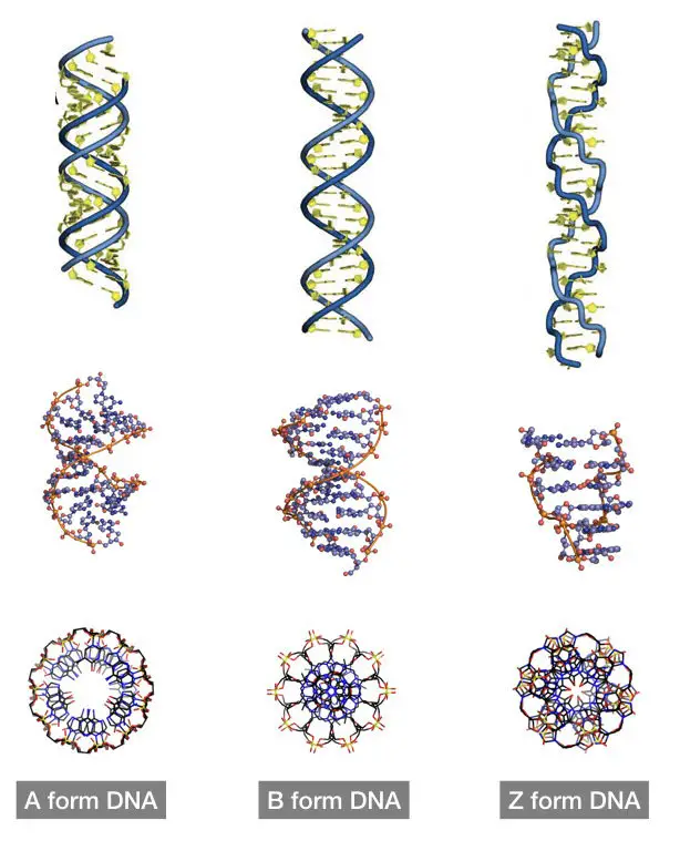
Types of DNA based on their structure:
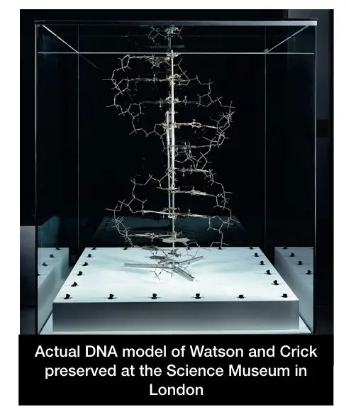
The DNA model demonstrated by Watson and Crick was the B-form DNA, however, it was originally discovered by Rosalind Franklin. It is the most prevalent form of DNA on earth, but, other DNA types like A, C and Z form DNA also exist. Let’s see different types of DNA depending on their topological features.
A form DNA:
A form DNA is right-handed in symmetry and highly similar to the B form DNA. The A-form (and B-form) DNA was discovered by Rosalind Franklin. A-form is shorter, more compact and smaller (approx. 20 to 25%) than the B form DNA and is present in the dehydration condition.
Due to the smaller twist angle which is titled around 20°, A-form DNA consists of ~11 base pairs per helix turn. The major and minor groove configurations also differ significantly. A major groove is deeper and narrow while the minor groove is wide and shallow.
The diameter of A-form DNA is 2.3 nm. It is slightly more than that of B-form DNA. Although the two strands are antiparallel, unlike the B-form DNA, A-form is not symmetrical which is because the glycosidic bonds are opposite to one another.
The A-form DNA does not appear in normal physiological conditions and only appears when humidity is too low. Interestingly findings also showed that DNA crystals are present in A-form. Conclusively, A-form DNA is not exclusively available for typical life forms.
B-form DNA:
B- form DNA is most abundant in nature, studied well and is the most prevalent type of deoxyribose nucleic acid. It is present in a normal physiological condition.
Rosalind Franklin discovered the B-form DNA using X-crystallography while the structure was chemically explained by Watson and Crick in 1953. Both the findings were groundbreaking discoveries in the history of science.
The B-form DNA is a right-handed helix having 10.5 bases per helix turn. The diameter of the helix is ~2.0 nm having a base pair title of -6°. The helix has two antiparallel strands which are asymmetrical.
The distance between two bases is 3.4Å. B-form DNA also consists of major and minor grooves; however, the major groove is wider and deeper compared to the major groove of A-form DNA. The B-form is shorter than the A-form DNA.
Z-form DNA:
The third active biological structure is Z-form DNA. It is unlike the A and B forms, the left-handed helix having a zigzag structure. The left-handed DNA was first reported by Robert Wells. However, the Z-form DNA was explained by Alexander Rich et al.
The typical Z form consists of 12 bases in a turn, 0.37 nm distance between two bases and 1.8nm width. It also has major and minor DNA grooves, however, the minor groove here is deeper and narrow.
| Attribute | A-form | B-form | C-form | Z-form |
| Helix sense | Right-hand | Right-hand | Right-hand | Left-hand |
| Base pairs per turn | 11 | 10 | 9.33 | 12 |
| Base pair tilt | 20° | -6° | 7° | |
| Rise/bp | 2.6Å | 3.4Å | 3.3Å | 3.6-3.8Å |
| Repeat unit | 1 bp | 1 bp | 1bp | 2 bp |
| Glycosyl bond conformation | Anti | Anti | Anti- pyrimidines; Syn: purines |
C-form DNA:
Yet another well-reported form of DNA is C-DNA. It was reported by Marvin in 1961. The C-DNA is less abundant and does not appear naturally in living beings. Interestingly, it appears only in the presence of certain ions like Mg2+ or Li+. It also appears in low humidity as well.
It consists of roughly 9.33 bases per helix turn. The sense of C-DNA is right-handed. Check out this comparative table to understand these points more accurately.
Types of DNA based on their topology:
Now let’s see different types of DNA based on their shape and topological properties. DNA topology is defined as the “twists and supercoiling that DNA contains.” Circular, linear and supercoiled DNA are three types of topological DNA forms.
Circular DNA:
The present DNA form is the most primitive and simplest form of DNA. It lacks any supercoiling. The ends are joined and lack twists and writhes. It is most prevalent in prokaryotic organisms. For example, plasmids in bacteria.
It is also present in eukaryotes as a sub-genome in the membrane-bounded organelles like mitochondria and chloroplast. Covalently closed circular DNA (cccDNA) is also one type of circular DNA, that appears only when a virus enters into the host cell.
Read more: What is rcDNA?- rcDNA vs cccDNA.
Linear DNA:
Linear DNA also lacks any supercoiling— twists and writhes. These are the smaller DNA fragments present in the sub-nuclear spaces and membrane-bounded organelles like chloroplast and mitochondria.
Some prokaryotes do have linear DNA but it is less abundant in nature, broadly.
Supercoiled DNA:
The most common form of DNA in nature is supercoiled DNA. Supercoiled DNA is made up of twists and writhes. Using proteins, the DNA coils and supercoils around itself and forms the most compact structure.
This helps the whole DNA to fit in the cell nucleus. Larger eukaryotic genomes supercoil to fit in the cell nucleus. Supercoiling mainly regulates gene expression by allowing and disallowing protein-DNA interactions.
Read more: Full Form of 10 Types of DNA.
Wrapping up:
DNA is our genetic material, henceforth, is so crucial for us to survive here on planet Earth. However, the activities in which the DNA is involved are more complex and very sensitive.
Mutation and alteration in gene expression are two faults in the process that can produce undesirable traits. Mutations commonly produce inherited genetic disorders while change in the expression of a gene may commonly result in cancer-like conditions.
Notedly, sometimes, these two may also give us some extraordinary survival skills.
Though B-form DNA is commonly found, other forms also appear in different physiological conditions. Fever evidence suggests the association of conformational changes of DNA with any genetic diseases.
So this is all about the basics of DNA. I hope you enjoyed this article. If so, please bookmark the page and share it.


Could you please put in the picture of how exactly DNA single unit looks like? I have been searching it a lot but seriously I didn’t get it. Please put the picture along with your definition, so that I can relate it with what is written.
Hello Ajitha.
The single unit of DNA is known as nucleotide which is a combination of nitrogenous base + phosphate + sugar. I had given its picture somewhere in this article. You can read our article: http://geneticeducation.co.in/nucleotides-vs-nucleosides/ to learn more