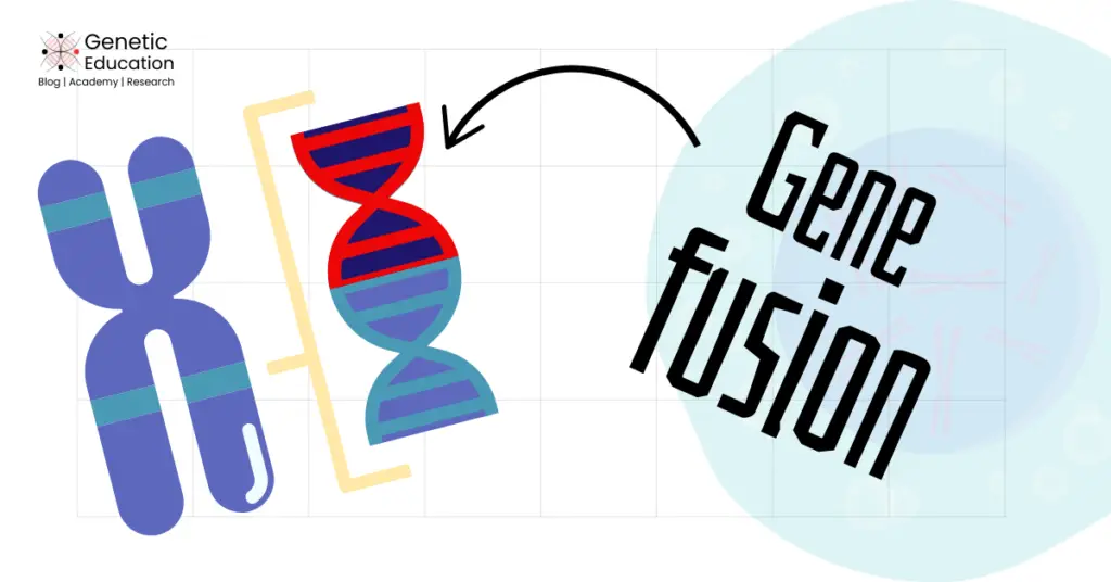
“When two independent genes combine because of Chromosomal translocations, Deletions, or Chromosomal inversions, they form a single hybrid gene. This phenomenon is known as Gene fusion.”
Cancer is genetic! Meaning that it originates through genetic mutations, either gene or chromosomal level alterations.
At the molecular level, it is recognized as an imbalance between proto-oncogene and tumor suppressor genes, with proto-oncogene mutations being the most common cause of cancer.
These proto-oncogenes convert into the mutant counterpart oncogenes through SNPs, deletions, duplications, gene amplification, translocation and gene fusion.
In this article, I will explain to you one of the most common events that occur in cancer— gene fusion- its definition, mechanism, types, consequences, and detection techniques.
Stay tuned.
Disclaimer: The content presented herein has been compiled from reputable, peer-reviewed sources and is presented in an easy to understand manner for better comprehension. A complete list of sources is provided after the article for reference.
Key Topics:
What is a Gene fusion?
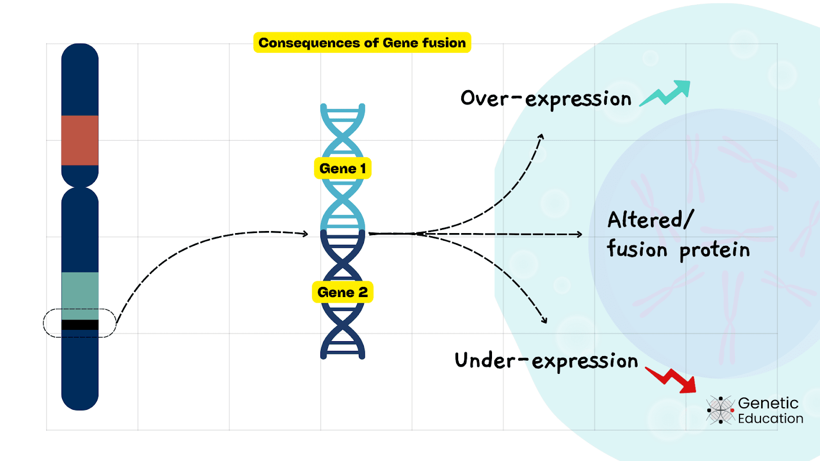
Gene fusion occurs when two genes are combined due to a random event. It is an important genetic event that often leads to genetic diversity. However, it also produces lethal conditions like cancer.
Over 30,000 gene fusions have been reported to date across all the chromosomes. Among these, 10,000 gene fusions have a clear role in cancer. Meaning that the majority of gene fusions have some positive effect on our genome.
Gene fusion has been a known evolutionary force for generating genetic diversity. The combination of two genes either suppresses an individual gene’s activity, overexpresses it, or produces a new type of protein.
In nature, gene fusion has significant importance for the origin of new genes, the evolution of protein domains & complex proteins, and the production of biologically important gene variants. This contributes to genomic diversity and complex genome evolution.
For instance, the immunoglobulin diversity, contributing to adaptive immunity, is developed through the fusion of V-(D)-J (variable, diverse, and joining) gene segments of the immunoglobulin gene.
V-(D)-J gene fusion occurred by the recombination event that produced different combinations (or types) of diverse antigen receptors. This gene fusion and production of novel gene variants have been an important genetic event for our immune system.
Several proteins have diverse biological functions that are contributed by different protein modules. Such complex proteins were evolved through the gene fusion process. For instance, the ABC transporter protein, the trpA & trpB gene fusion and the antibiotic-resistant genes evolved through gene fusion.
A candidate gene in vertebrate limb development, the HOX gene is also evolved through gene fusion. Similar to HOX, in Drosophila, the novel body pattern protein was developed by Antp-Scr gene fusion.
Several examples of gene fusion and their biological significance are given in the table below.
| Event | Gene fusion | Organism | Importance |
| Antibiotic Resistance | Antibiotic resistance gene fusion | Bacteria | Resistance to antibiotics |
| Environmental Adaptation | Light-sensing gene fusion | Cyanobacteria | Enhanced photosynthesis efficiency |
| Yeast Mating Type Switching | MATa and MATα | Saccharomyces cerevisiae (Yeast) | Enables yeast to switch mating types for better adaptation |
| Antifreeze Protein Evolution | AFPIII fusion from repetitive sequences | Antarctic Fish | Prevents ice crystal formation in blood, aiding survival in freezing waters |
| Opsin Gene Fusion in Vision Evolution | RH1 and RH2 | vertebrates | Improved color vision and light sensitivity in mammals and birds |
Gene fusion as an evolutionary force that helped organisms to develop novel proteins and traits to survive on earth. There are thousands of examples out there in nature, that show how important gene fusion events are; however, it isn’t always the case!
Unwanted gene fusion events produce adverse health-related issues and lead to cancer-like conditions.
Definition
Gene fusion is a genetic event that occurs by chromosomal rearrangements like translocation, inversion or deletion, leading to chimeric or altered protein production. Gene fusion is commonly observed in cancer.
Gene fusion in cancer:
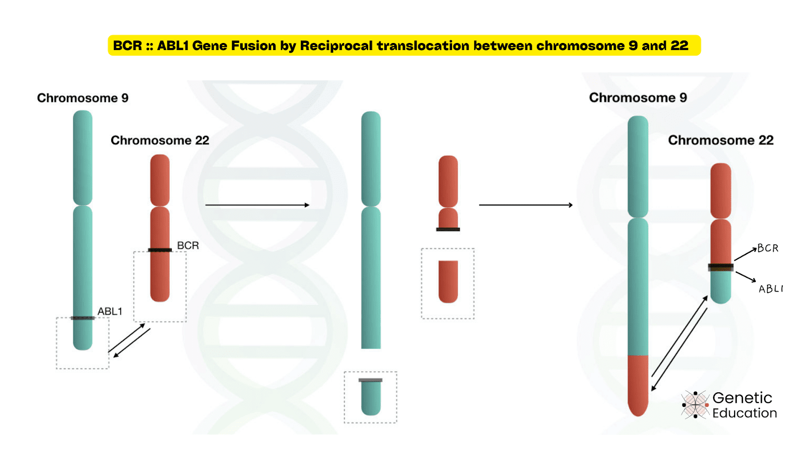
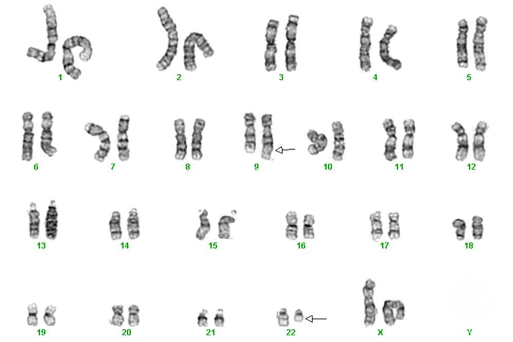
Gene fusion is also commonly reported in cancer. The role of gene fusion in cancer was described in 1980. It was first identified in chronic myeloid leukemia, between chromosome 9 and 22, the condition known as the Philadelphia chromosome.
In 1973, Janet Rawley, a scientist from Chicago, described that the Philadelphia chromosome had originated through a translocation between chromosomes 9 and 22.
Initially, it was thought that the Philadelphia chromosome originated by simple deletions. In 1980, several researchers found that the translocation in the Philadelphia chromosome resulted in the fusion of the genes BCR and ABL1, which led to the formation of the BCR-ABL1 fusion gene. This gene produces a protein that can induce chronic myeloid leukemia.
Various mechanisms of gene fusion:
Various types of chromosomal instabilities such as translocations, deletions or inversions lead to gene fusions. In addition, errors in DNA repair and replication and activation of mobile genetic elements also lead to gene fusion.
Rearrangements at the chromosomal level occur due to several natural as well as in vitro processes, further leading to gene rearrangements, and in some cases, two independent genes are combined, which results in a fusion gene.
Gene fusion because of chromosomal rearrangements occurs as a result of translocations, inversions, and interstitial deletions. A few gene fusion examples are explained here.
Chromosomal translocations:
During cell division, a piece of chromosome breaks and rejoins to a different chromosome; this phenomenon is called a translocation. Common types of translocations are balanced, unbalanced, reciprocal, and robertsonian translocations.
Among all these, reciprocal translocation results in gene fusion without losing material.
Reciprocal translocations, where two different chromosomes (non-homologous) exchange pieces with each other, mainly result in gene fusion. The most common example is chronic myeloid leukemia, which results from a reciprocal translocation between chromosomes 9 and 22.
Here, the ABL1 gene from chromosome 9 breaks and joins with the BCR gene on chromosome 22. Chromosome 22 with BCR-ABL fusion gene is called the Philadelphia chromosome. The BCR::ABL1 fusion gene is found in most people with chronic myelogenous leukemia.
Another example is ETV6-NTRK3 gene fusion. The ETV6 gene is located on chromosome 12 and the NTRK3 gene is located on chromosome 15. Translocations between these two chromosomes result in ETV6-NTRK3 gene fusion.
In this case, the ETV6 promoter is more active than the NTRK3 promoter, so the fusion gene is highly expressed in certain tissues, resulting in Lung carcinoids, Congenital fibrosarcomas, and Pilomyxoid astrocytomas.
Chromosome inversions:
Chromosome inversion also causes gene fusion and results in tumor formation.
Chromosome inversion is when a piece of chromosome breaks and rotates 180 degrees and rejoins into the same chromosome. One of the most common examples is the FGFR1::PLAG1 fusion gene, which results from pericentric inversion in chromosome 8.
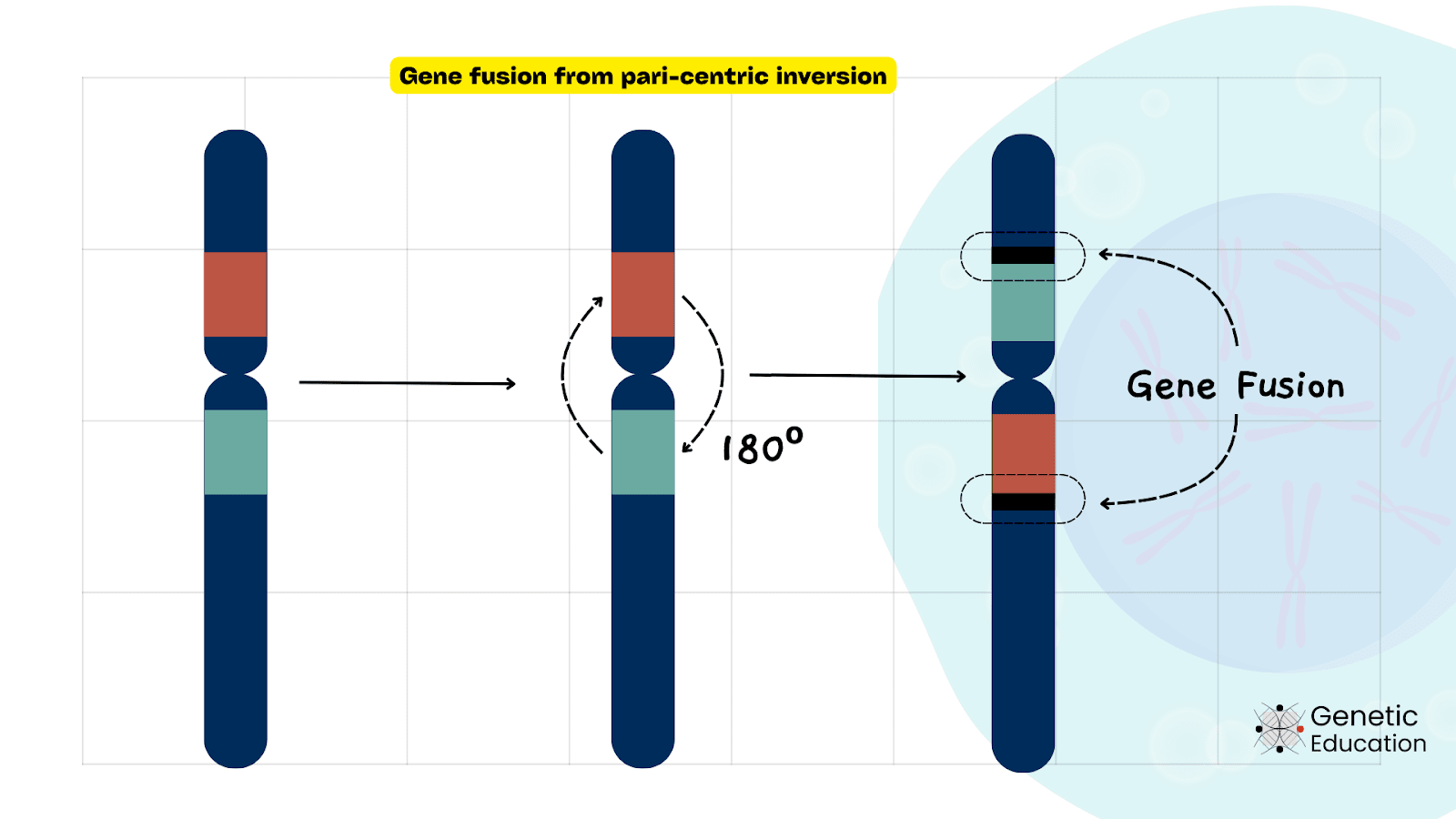
This inversion results in mitotically unstable chromosome structures such as ring chromosomes and dicentric chromosomes. This type of gene fusion is responsible for certain pleomorphic adenomas and myoepithelial carcinomas.
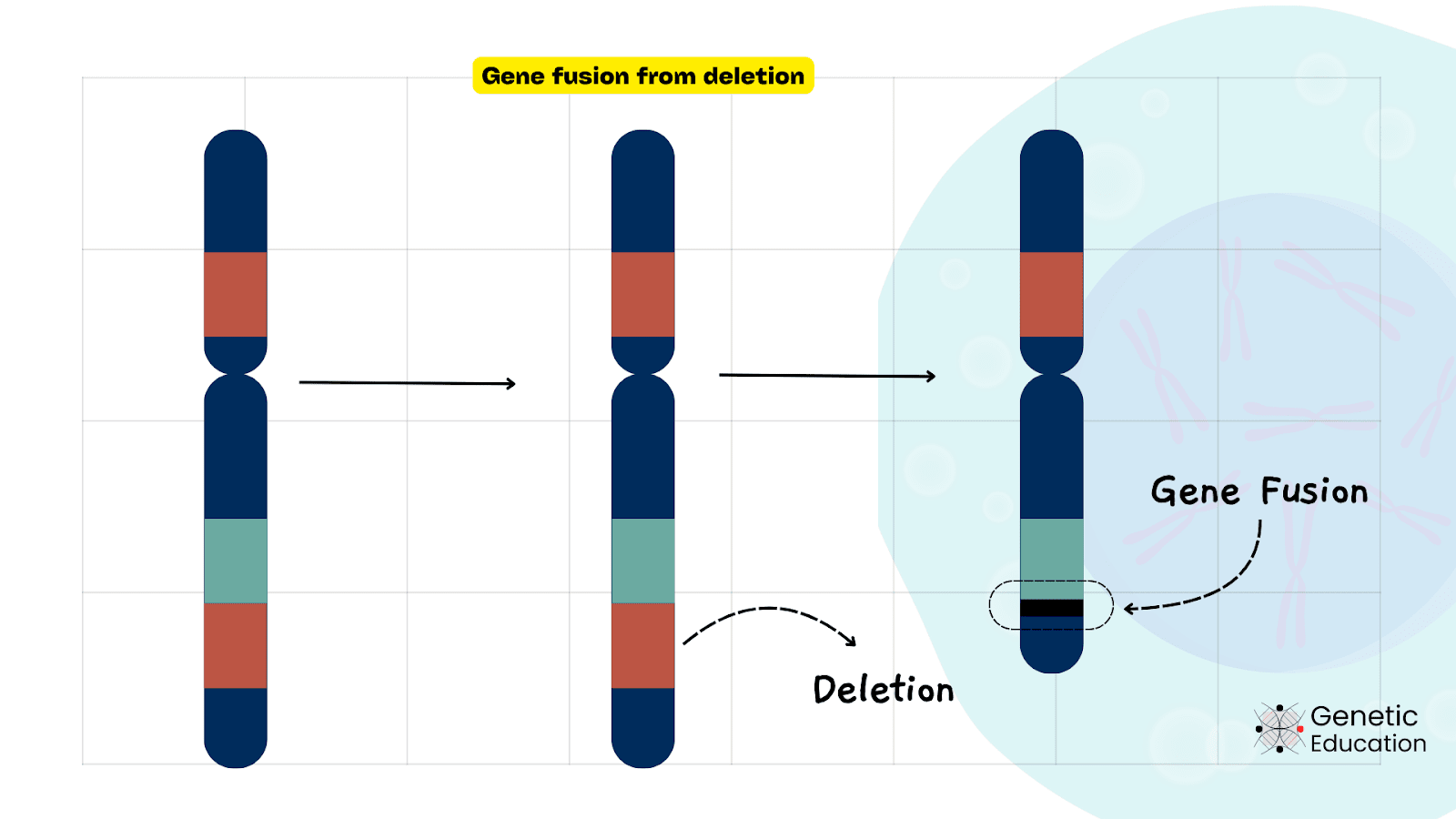
Chromosome deletions:
Chromosome deletion is when a part of the chromosome is deleted. This results in structural chromosome rearrangements, bringing two separate genes together and forming a fusion gene.
One of the most common examples of gene fusion by deletion is the PAX7-FOXO1 fusion gene. In this, part of chromosome 13 breaks and results in the PAX7-FOXO1 fusion gene, which is responsible for rhabdomyosarcoma.
Trans-Splicing of RNA:
During RNA processing stages after transcription, two separate RNA transcripts from different genes are spliced together, which is called trans-splicing of RNA, resulting in gene fusion at the RNA level.
This type of RNA splicing produces the abnormal protein, which can cause certain types of cancer.
One of the most common examples is the JAZF1-JJAZ1 gene fusion, which is detected in 50% of endometrial stromal sarcomas (ESSs) and almost all benign endometrial stromal nodules (ESNs).
Transcription-derived gene fusion (TDGFs):
During transcription, the adjacent genes produce a novel transcript, which is called Transcription-derived gene fusion. One of the most common examples is the LC45A3-ELK4 fusion found in prostate cancer.
Types of gene fusion:
Depending on the number of genes involved in the fusion, they are categorised into double or triple gene fusion.
Double gene fusion:
When a piece of the chromosome with one gene breaks and reattaches with another gene, which creates a new hybrid gene with combined functions, it is called double gene fusion.
Examples of double gene fusion include TMPRSS2-ERG fusion in prostate cancer, FGFR3-TACC3 fusion in glioblastoma, EML4-ALK fusion in lung cancer and BCR-ABL fusion in leukemia.
Triple gene fusion:
Triple gene fusion happens through multiple chromosomal breaks where pieces of multiple genes are joined together. Examples of triple gene fusion include ETV6-AML1-NPM1 fusion in myeloid leukemia, TMPRSS2-ERG-PTEN fusion in prostate cancer and ALK-EML4-NPM1 fusion in lung cancer.
Consequences of Gene Fusion
Oncogenic gene fusion:
Gene fusion often produces an abnormal protein, which in the majority of the cases observed in various types of cancer, and also contributes to the tumorigenesis.
Common examples are BCR-ABL1 fusion in chronic myeloid leukemia, ETV6-RUNX1 fusion in acute lymphoblastic leukemia, TMPRSS2-ERG fusion in prostate cancer and EML4-ALK in lung cancer.
Now, the gene fusion in cancer results in oncogene activation. The fusion of two or more genes mutates the proto-oncogene. This causes overexpression of the protein associated with the proto-oncogene and results in cancer.
Genetic disorders:
When parts of two or more genes are joined together, it can also result in genetic disorders. Familial Down syndrome is one of the most common examples of genetic disorders caused by gene fusion. This fusion is the result of translocation between non-homologous chromosomes 21 and 14.
Detection methods for gene fusion:
Gene fusions have been detected using Karyotyping, Southern blot analysis, fluorescence in situ hybridisation (FISH), polymerase chain reaction (PCR) and next-generation sequencing.
Southern Blot analysis:
A conventional and old-fashioned molecular approach for detecting gene fusions at the DNA level that identifies certain restriction fragments hybridized with labelled probes.
In the Southern blot, particularly for gene fusion analysis, the genomic DNA is digested with restriction enzymes, separated by gel electrophoresis, transferred to a membrane, and hybridized with a labelled probe to target the fusion breakpoint.
It is useful to detect gene fusions at the DNA level, however, it has several drawbacks. It requires radioactive probes (not safe to work with!) and a large amount of starting material or tissue.
Low abundant gene fusions can’t be effectively studied, which makes it a least sensitive technique. It is also time-consuming and labour-intensive.
Karyotyping:
Karyotyping is a cytogenetic technique used to detect chromosome rearrangements such as chromosomal translocations, inversions, and deletions. Abnormal chromosomes such as the Philadelphia Chromosome can be detected by karyotyping.
Since karyotyping can detect only large chromosomal changes, it can not be used to detect the smaller gene fusions. To know more on karyotyping, you can click the link and read the complete article.
Fluorescence In Situ Hybridization (FISH):
FISH (Fluorescence In Situ Hybridization) is a molecular cytogenetic technique used in the detection of various chromosomal abnormalities. FISH is also useful in the detection of gene fusion by using fluorescently labeled DNA probes.
In this technique, probes are designed in a way that they bind with the gene fusion region, provide fluorescence signals, and are detected using fluorescence microscopy. For instance, in the case of di-color FISH, two different FISH probes are used for two different genes involved in the fusion.
For example, in the case of BCR-ABL gene fusion FISH, red colored and green colored probes are used for BCR and ABL genes, respectively. The fusion event can be effectively tracked using two-color FISH.
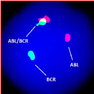
FISH is fast, accurate and sensitive enough to detect cancer rearrangements. Thus, FISH is widely used in the detection of various cancers, including cancers caused by various gene fusions.
Polymerase Chain Reaction (PCR) and Reverse Transcription PCR (RT-PCR):
Conventional PCR, qPCR, and RT-PCR, all three variants have been extensively used to detect known gene fusions at the DNA level. Using the sequence-specific primers, the gene fusion can be detected.
The quantitative reverse-transcriptase PCR has been used to determine the gene expression of the fusion genes. This detection technique has high specificity and sensitivity, but it can be used only when fusion genes and breakpoints are known.
Recently developed digital PCR or droplet digital PCR has also been used for the accurate quantification of rare gene fusions.
DNA Sequencing:
Sanger sequencing is a gold-standard method in diagnosis. It is particularly utilized for cancer diagnosis, screening, prognosis, and treatment outcome. Gene fusions can also be determined using Sanger sequencing. Importantly, it provides sequence-level information.
Another high throughput sequencing technique, next-generation sequencing, is also used for gene fusion analysis. This technique can identify novel gene fusions, trans-splicing of RNA and transcription-mediated gene fusion.
NGS is expensive and requires a sophisticated computational and bioinformatics setup.
Wrapping Up:
Gene fusion has pivotal evolutionary importance. Although it isn’t as aggressive as gene amplification, it causes various types of cancer. Notably, it isn’t inherited and can’t be transmitted in the majority of cases.
Our cells’ natural DNA repair mechanisms can repair many mutations. Gene fusions belong to the category of mutations that can’t be repaired effectively.
Techniques like karyotyping, FISH, PCR variants, and DNA sequencing are extensively used for the diagnosis of cancer and for studying and identifying gene fusions.
I hope you like this article. Do share it and subscribe to Genetic Education.
References:
Wikipedia contributors. (2024, August 5). Fusion gene. Wikipedia. https://en.wikipedia.org/wiki/Fusion_gene.
Translocation.Genome.gov.https://www.genome.gov/genetics-glossary/Translocation
NCI Dictionary of Cancer Terms. Cancer.gov. https://www.cancer.gov/publications/dictionaries/cancer-terms/def/bcrabl1-fusion-gene
Stenman, G., Fehr, A., Skálová, A., Poorten, V. V., Hellquist, H., Mikkelsen, L. H., Saba, N. F., Guntinas-Lichius, O., Chiesa-Estomba, C. M., Andersson, M. K., & Ferlito, A. (2022). Chromosome translocations, gene fusions, and their molecular consequences in pleomorphic salivary gland adenomas. Biomedicines, 10(8), 1970. https://doi.org/10.3390/biomedicines10081970
Li, H., Wang, J., Ma, X., & Sklar, J. (2009). Gene fusions and RNA trans-splicing in normal and neoplastic human cells. Cell Cycle, 8(2), 218–222. https://doi.org/10.4161/cc.8.2.7358
McCartney, A. M., Hyland, E. M., Cormican, P., Moran, R. J., Webb, A. E., Lee, K. D., Hernandez-Rodriguez, J., Prado-Martinez, J., Creevey, C. J., Aspden, J. L., McInerney, J. O., Marques-Bonet, T., & O’Connell, M. J. (2019). Gene Fusions Derived by Transcriptional Readthrough are Driven by Segmental Duplication in Humans. Genome Biology and Evolution, 11(9), 2678–2690. https://doi.org/10.1093/gbe/evz163
Understanding Chromosome Disorders | Thisability. (n.d.). Thisability. https://www.thisability.org/understanding-chromosome-disorders.
Zhou, Y., Zhang, C., Zhang, L. et al. Gene fusion as an important mechanism to generate new genes in the genus Oryza. Genome Biol 23, 130 (2022).

