“Restriction Digestion is a common and conventional genotyping technique. Learn about the process and protocol of restriction digestion and other related information in this article.”
Restriction digestion is a targeted DNA fragmentation technique. It is widely used in genotyping, popularly known as RFLP (Restriction Fragment Length Polymorphism). It includes subsidiary techniques like PCR and gel electrophoresis for DNA amplification and analysis, respectively.
Despite having many high-throughput and robust genotyping techniques, restriction digestion is still popular because of its high accuracy rate. However, the success rate depends on how you design the experiment and utilize your protocol.
Thus, a well-established and keenly standardized restriction digestion protocol is needed. But before discussing the protocol, you first have to understand the concept of restriction digestion.
In this article, I will surely give you my standard restriction digestion protocol but before that let’s understand the concept, mechanism, process, advantages, limitations and applications of restriction digestion.
Stay tuned.
Disclaimer: Information provided in this article is collected from peer-reviewed resources and re-presented in an understandable language. All the sources are enlisted at the end of the article.
Key Topics:
What is Restriction Digestion?
Restriction Digestion is a process to cleave or digest a DNA sequence at a particular recognition site using a restriction endonuclease. It’s a widely used molecular genetic tool for various experiments and purposes.
Smith, Thomas Kelly and Kent Wilcox identified the very first restriction endonuclease in 1970. However, W. Arber introduced the term ‘endonuclease’ in 1960. Smith, Thomas Kelly and Kent Wilcox were awarded the Nobel Prize in 1978 for the present findings.
Interestingly, the mechanism of restriction digestion and the concept of endonucleases were first explained by Salvador Luria and Giuseppe Bertani during their experiment on bacteria— around 1950.
It was only after the 1980s, that scientists used the present mechanism for genetic engineering and genetic research. At present, restriction digestion is used in techniques like DNA sequencing, Microarray, gene editing (CRISPR-CAS9) and genetic manipulation.
Restriction endonucleases govern the process of restriction digestion. They are enzymes found in bacteria as a component of their defense system. They help bacteria against invading phages and viruses.
We have explained the concept of restriction enzymes and the 10 most common enzymes in this article: What are Restriction Enzymes? Top 10 Common Restriction Enzymes Used in Genetic Engineering.
Put simply, an endonuclease cleaves any DNA using its recognition site, which is a known sequence for an endonuclease enzyme. Scientists utilized the present concept to study sequence alteration, thereby genotyping.
Note:
Mechanism:
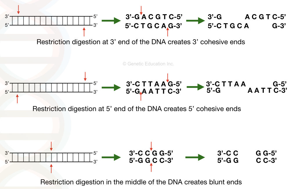
The restriction digestion process is catalyzed by a restriction endonuclease enzyme. The enzyme initiates catalytic activity only when it finds its recognition site. Often known as the restriction site, a recognition site is a known sequence location for an enzyme on a template DNA. For example– The recognition site for EcoR1 is GAATTC.
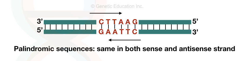
A recognition site is 4 to 6 nucleotides long, palindromic sequences, majorly. Palindromic sequences are short nucleotide sequences that remain the same when read from 3′ to 5′ or 5′ to 3′ directions.
To know more, read this article: What is a Palindromic Sequence?
Now, after that, the enzyme settles on the dsDNA (on its recognition site) and starts catalyzing the reaction using its catalytic core domain. It starts cutting the dsDNA from both strands and by disrupting the phosphodiester bonds.
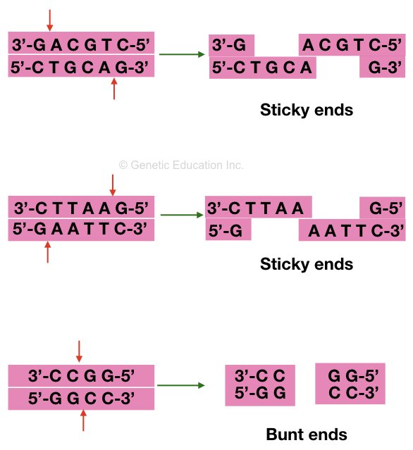
The catalytic action can produce two types of ends after cutting. One, sticky or cohesive ends and blunt ends.
Sticky ends have a few nucleotide overhangs and are generated when the enzyme cleaves at two different locations on its recognition site (on a dsDNA). Blunt ends are generated when the enzyme cuts at the same location in the middle of the recognition site.
Carefully observe the image.
Now based on the location and the number of recognition sites on a target DNA, different numbers of fragments with varied lengths are obtained. Note that as the number of recognition sites increases, the length of each fragment decreases.
For example, for one, two and three recognition sites we will obtain two, three and four DNA fragments, respectively.
Process:
The process of restriction digestion is not complicated, we can say but is a bit tedious as it involves wet and dry lab work. First, we have to choose the sequence, an enzyme, order it and then we have to perform the experiment.
Let’s understand the process step-by-step.
Selection of Target Sequence:
Target selection is the very first step and is crucial. Here, we first have to select a gene or sequence which we want to study. If we want to study a particular mutation like— SNP, insertion or deletion, we have to isolate the FASTA sequence format and locate our mutation in the sequence.
Note that if you are interested in a particular location, you can trim the sequence and use only the target part, however, it should be at least 1000 nucleotides long.
Selection of restriction endonuclease:
Now in the next step, we need to select a restriction enzyme for our experiment. Manually, it’s not possible to remember all the REs, their recognition sequence and their locations. So we use the tool, for example— NEB cutter.
What we have to do is, paste our sequence in the NEB cutter and it will give us the list of all the REases. Depending on the location of our mutation or SNP, we can choose a particular type of REase.
Now, we need to order the REase.
DNA isolation:
In the next step, we need to isolate DNA from the sample. DNA isolation is a simple process. You can use a manual protocol or any ready-to-use kit for DNA isolation.
PCR amplification
It’s important to know that there are millions of recognition sites present in the host genome, for a particular REase, that we selected. So to avoid result bias, what we do is, only amplify the sequence or gene which contains the recognition site only once or twice in the sequence.
To do so, we need to prepare the sequence-specific primers and amplify the sequence using the PCR. PCR amplification generates millions of copies of that particular fragment, and gives us more flexibility for results and analysis.
PCR results can be validated on agarose gel electrophoresis.
Restriction digestion
In the next step, we have to prepare a restriction digestion reaction. Here we add a digestion buffer, our PCR amplicon and restriction endonuclease and allow it to incubate. The enzyme cleaves all the amplicons wherever it finds its recognition site.
Results analysis:
After completion of incubation, the sample is run on an agarose gel electrophoresis to visualize, study and interpret the results.
Isn’t that a simple process?
Protocol for Restriction Digestion:
Requirements:
1.5 and 2 ml Eppendorf tubes, PCR master mix kit, restriction digestion kit, pipette, nuclease-free water, template DNA.
PCR Reaction preparation:
- Isolate high-quality DNA and use it for restriction digestion.
- Prepare a PCR reaction shown in the table below.
| Component | Concentration | volume |
| PCR master mix | 200 µM | 4 µL |
| Forward primer | 10 pM | 1 µL |
| Reverse primer | 10 pM | 1 µL |
| PCR buffer | 1X | 10.5 µL |
| Taq DNA polymerase | 1 U | 1.5 µL |
| Template DNA | 30 to 50 ng | 3 µL |
| Water | – | 4 µL |
| Total | 25 µL |
- Prepare PCR reaction using this sequence.
- Add 200 µM (4µL) dNTPs mix.
- Add 10 pM (1µL) forward and reverse primers in the reaction.
- Add 1U (1.5 µL) Taq DNA polymerase.
- Add 30 to 50 nG (3µL) template DNA.
- Add 1X (10.5µL) PCR buffer.
- Make a final volume of the reaction- 25 µL using the nuclease-free water.
- Put the reaction tube in the thermal cycler as per the PCR reaction conditions given in the table below.
| PCR step | Initial denaturation | Denaturation | Annealing | Extension | Final extension |
| Temperature | 94ºC | 94ºC | 50 to 65ºC | 72ºC | 72ºC |
| Time | 5 to 7 minutes | 45sec | 45sec | 45sec | 5 to 7 minutes |
| Cycles | 30 to 35 cycles |
Run the PCR amplicons on 2% agarose gel electrophoresis.
Restriction digestion reaction preparation:
Now prepare a restriction digestion reaction using the table given below.
| Reagent | volume |
| REase EcoRI (10 unit/μl) | 1 μl |
| 10X buffer (REase buffer provided by the supplier) | 3μl |
| PCR product | 10μl |
| Nuclease-free water | 16μl |
| Final volume | 30μl |
- To prepare a restriction digestion reaction first add 10μl PCR product in a sterile tube.
- Add 1μl restriction enzyme (10 units/μl).
- Add 3μl (10X) reaction buffer.
- Make up the final volume up to 30μl with the help of nuclease-free water.
- Gently vortex mix the sample and incubate the sample at 37ºC overnight or at 60ºC for 1 to 2 hours (depending on the type of enzyme we have).
- After incubation, run the reaction on 3% agarose gel for 1 to 1.5 hours.
- Note: Heat inactivation is required for some enzymes to stop their activity. To do this, after incubation, heat the sample for 10 to 20 minutes at a higher temperature (80ºC).
Advantages:
- The present molecular genetic tool is precise. It comes with ultra-high accuracy as it uses a recognition site to cleave the DNA. The REase cuts exactly where its recognition site is located.
- The experimental setup for restriction is very simple. Add an enzyme to the sample and incubate it, that’s it.
- The experiment is relatively cheaper.
- It doesn’t require high-end instrumentation for analysis. A simple agarose gel electrophoresis is enough to study the results.
Limitations:
The present technique has unmatched applications, even in the present time but also has serious limitations. Here are some.
- It requires sequence information to prepare the experiment and thus, unknown sequence variations can’t be determined. In addition, some sequences or genes that lack recognition sites or contain multiple recognition sites, create difficulties in the experiment.
- It’s a time-consuming process.
- It is highly contaminant-prone. The chances of cross-contamination are very high here.
- Restriction digestion doesn’t work for methylated DNA sequences.
- Overlapping recognition sites make experiment difficult in the case of multi-enzyme digestion.
- The dependency on the recognition site restricts its applications.
- Incomplete digestion under inadequate conditions is also a major problem with the present technique.
- A few types of enzymes may unintentionally cut sequences other than their recognition site when reaction conditions are compromised.
- It needs wet and dry lab work and thus lab personnel has to have thorough knowledge regarding computational and lab work.
- The present technique has limited throughput.
- Separate experiment setup and design are required for every experiment.
Applications:
Despite having the above-listed limitations, restriction digestion is widely used in genetic research, genetic engineering, gene therapy and gene manipulation experiments.
Genotyping:
Restriction digestion is the pivotal technique for genotyping. Known sequence SNPs, insertions, deletions or CVNs can be determined. For example, in our COMT gene study, we selected one candidate mutation from the COMT gene and digested it with NIaIII. You can read that research paper here: Role of TH and COMT gene mental retardation.
Recombinant DNA technology:
The present technique is extensively used in recombinant DNA technology and genetic engineering experiments, to prepare recombinant plasmids. To insert the gene of interest into the plasmid, the plasmid DNA and GOI are digested using the same type of REase.
Using a T4 DNA ligase the sticky ends of the GOI and plasmid have been sealed.
Restriction mapping:
The present technique is often utilized for restriction mapping as well. In this process, a whole map of the chromosome or genomic DNA is prepared by digesting the sample with the sample type of REase.
Marker assistant studies:
Restriction digestion is the primary technique for markers like RFLP (Restriction Fragment Length Polymorphism) here, various polymorphic regions or sequence alterations can be studied.
Discriminate between homozygous and heterozygous:
It is also employed for homozygous and heterozygous discrimination and related studies. Restriction digestion is a gold standard technique for this purpose.
Genomic library preparation:
It is also used in techniques like DNA sequencing or microarray for preparing fragment libraries. Fragment libraries are prepared by digesting the sample with a REase and ligating it with known sequence adaptors.
Wrapping up:
Restriction digestion is a crucial molecular genetic tool, especially for genotyping. It’s a super easy, cheap and reliable option for small-scale research. I personally used this technique in many of my research projects.
I personally like it, trust me the results are so reliable and trustworthy. However, at the same time, it’s a time consuming and low throughput technique. That’s it for the present article. I will write more articles on this topic and update them here.
I hope this article will help you in your learning. Do share the article, comment below if you like it and bookmark the page.
Sources:
Smith DR. Restriction endonuclease digestion of DNA. Methods Mol Biol. 1993;18:427-31. doi: 10.1385/0-89603-245-0:427. PMID: 21390690.
Loenen WA, Dryden DT, Raleigh EA, Wilson GG, Murray NE. Highlights of the DNA cutters: a short history of the restriction enzymes. Nucleic Acids Res. 2014 Jan;42(1):3-19. doi: 10.1093/nar/gkt990. Epub 2013 Oct 18. PMID: 24141096; PMCID: PMC3874209.

