“The chromatin is a bead-on-string like structure made up of a complex network of histone proteins and DNA and helps nucleic acid to fix inside a cell.”
DNA is a mysterious thing, as we understand it, its mystery becomes more and more mysterious. DNA is a complex molecule understood well in 1953. We have now sufficient information regarding DNA.
It is an information storage and transport unit means it inherited information.
It replicates to become double and can also repair damaged DNAs. Some special type of functional sequences known as a gene makes proteins.
All the proteins are formed from the genes and their expression is regulated through the DNA as well.
Changes occur in DNA and that leads to the evolution of new phenotypes and traits in nature. That is why DNA is so important for us.
DNA is longer than we think. As per some evidence, if we unpack the DNA of all cells and stretch it, we can go to the moon and even come back. The DNA thread of a single cell is approximately 2 feet long.
So to make it fit inside a cell, it should be arranged properly. The process known as DNA packaging helps DNA to fit inside a cell. Through the various level of organization, DNA makes it possible to arrange in a cell and replicate.
Related article: DNA: Definition, Structure, Function, Evidence and Types.
In the present article, our major talk will be on chromatin, a special type of arrangement that helps to make chromosomes. We will also discuss its functions.
Key Topics:
What is chromatin?
Before going into the present topic I strongly recommend reading our previous article on DNA packaging. This article has all the information on different stages of DNA compacting. Read it here: DNA packaging in eukaryotes.
Prokaryotic are simple and single-cell organisms having a simple DNA arrangement. On the other hand, the eukaryotic organisms are multicellular having supercoiled DNA.
Due to the complex arrangement, our DNA (eukaryotic DNA) is different from the prokaryotes. DNA of eukaryotes are arranged on chromosomes, while the prokaryotes have a single circular chromosome on which their genetic material is located. Learn more on this topic: Prokaryotic DNA vs Eukaryotic DNA.
The complex network of DNA, its supercoiling, and compactness make it unique and different from prokaryotic DNA.
Nucleosome, chromatin, chromatid, and chromosomes are different stages of arrangements.
Definition:
“A chromatin is a complex structure of histones and DNA that makes it possible to fit DNA in a cell by forming a chromosome.”
Structure:
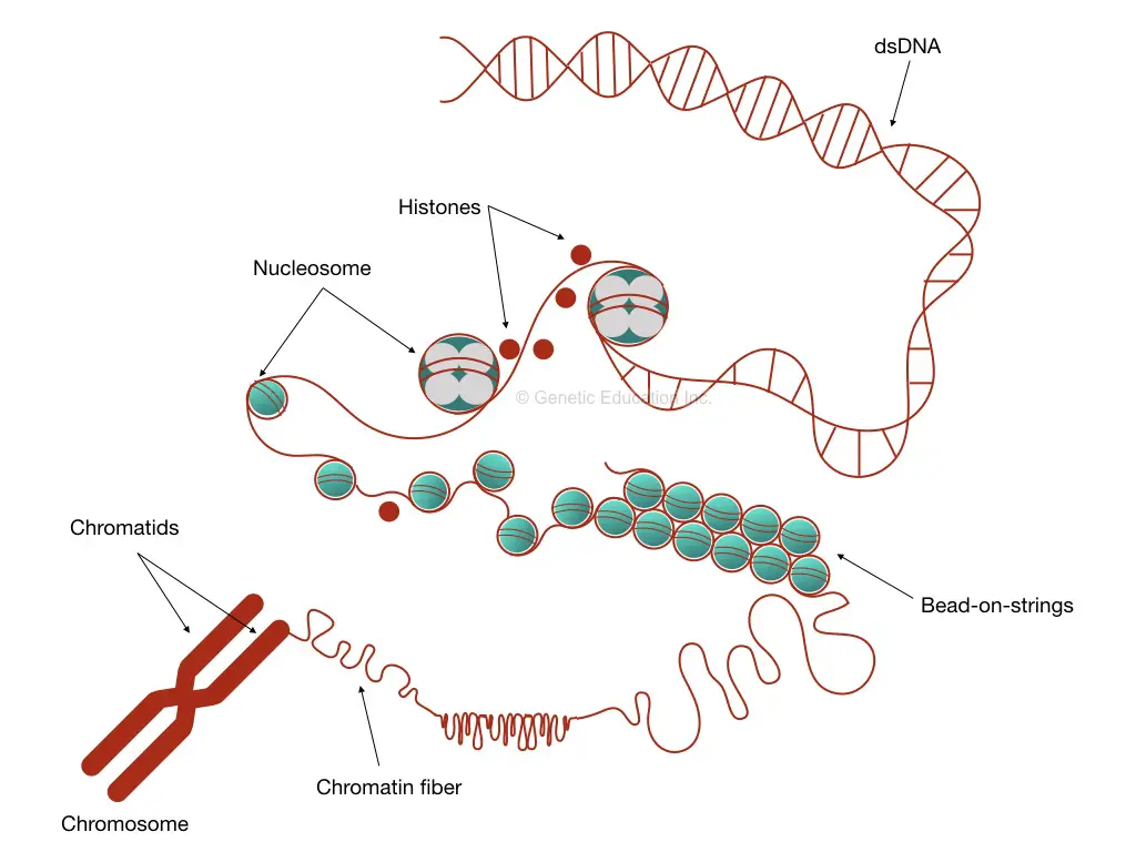
So let’s start with the DNA itself. Two strands of DNA wind with one another and creates a DNA helix. It further interacts with other molecules, coils on one another, and creates a supercoiled form of DNA.
Information: supercoiling is a characteristic of the eukaryotic genome.
The protein that is involved in this process is known as histones.
In the path of chromatination (formation of chromatin), nucleosome structure forms first. A nucleosome is an arrangement of 147bp DNA wrapped around the octa-core of histones.
The histone octamers are the two units of histone H2A, H2B, H3 and H4. Thus it clearly indicates that chromatin has twice as much nuclear protein as DNA.
As we said, 4 different types of proteins as commonly involved in the primary level of the organization, its mass is equal to the DNA of the nucleus. Lysine and arginine are two common types of amino acids present in histones that facilitate the binding of the protein to DNA.
Information: more than 1000 different non-histone and histone proteins are involved in the formation of chromatin, chromosome and to perform replication and transcription, and gene expression.
Notably, histone is not observed in prokaryotes but it is believed that similar proteins like it might be involved in the packaging of prokaryotic DNA.
Interestingly, the chromatin subunits are also formed by the linker protein that joins two DNA known as H1 histone. The subunits are known as chromatosomes which have 166bp of wrapped DNA.
The chromatosomes with the 80 base pair linker DNA forms a thread of 10nm fiber that will further organize to form a 30nm fiber. Due to less published data or 30nm its structure is not studied well.
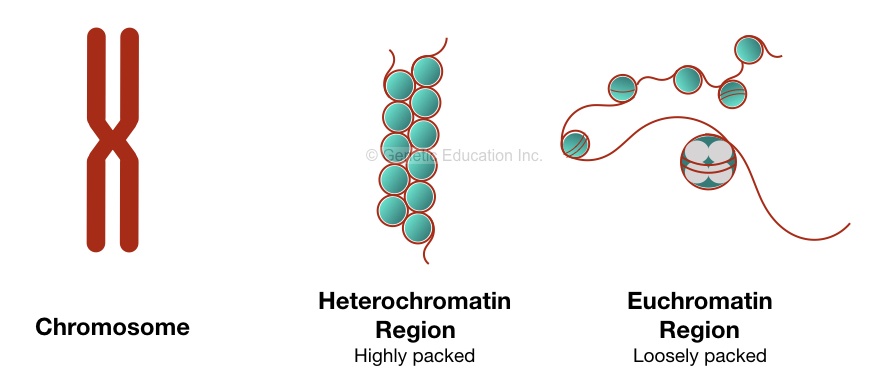
We are not going into the detail here, the molecular structure of chromatin is complicated and yet not understood well. Based on how compactly the DNA is arranged! The chromatin is divided into two parts; euchromatin regions and heterochromatin regions.
Euchromatin regions:
The concept of chromatin condensation is to allow and disallow the transcription.
The euchromatin regions are loosely packed which allows enzymes to work. Enzymes can settle on DNA of loosely packed DNA and facilitate transcription.
The RNA polymerase can settle on euchromatin regions and forms the mRNA transcript from the DNA and thus a protein is formed from it.
That is why the Euchromatin regions are too important for a cell to survive. Usually, Euchromatin is observed during the interphase of cell division. The interphase is a condition in which the cell is not dividing but other DNA activities like replication and transcription happen.
As we said, loosely packed chromatin allows various enzymes to catalyze the reaction. The euchromatin region stains light blue by Giemsa stain due to fewer protein parts.
Heterochromatin regions:
Contrary to the euchromatin regions, the heterochromatin regions are compactly and tightly packed regions that block DNA metabolism. Enzymes like RNA polymerase can’t find binding sites on DNA due to tight wrapping. And due to that mRNA transcript can’t be formed.
Usually, the metaphase chromosomes are tightly packed heterochromatin which means it doesn’t allow any other activity for enzymes. The metaphase chromosomes are segregated during metaphase into two daughter cells.
Note: Not all the DNA during the interphase are euchromatin and loose, some DNA is heterochromatin which regulates gene expression.
Functions of chromatin:
There are several reasons why DNA packs, loosen and unpack. The entire mechanism regulates and facilitates several DNA metabolic activities.
Replication:
Replication is a process of doubling of DNA. while replication, tension occurs on the rest of the double-stranded DNA. An enzyme known as DNA topoisomerase helps to release tension. The loosely arranged DNA is more prone to replication.
Transcription and gene expression:
Transcription is a process to form mRNA by RNA polymerase.
The key function of chromatin remodeling is to give access to gene expression and transcription. We have already discussed it. Chromatin is a kind of signal for transcription.
Related article: “Transcription And Translation” A Brief Overview.
DNA repair:
DNA repair is as important as DNA replication. During harsh conditions DNA damages. Chromatin is a dynamic and flexible structure that also allows DNA repair as soon as possible.
At a site of damage, chromatin changes its shape, loosens, and allows enzyme activity. The polymerase identifies the site, settles on it and repairs the DNA by adding the nucleotides to the damage site.
The process known as chromatin remodeling mediated by the PARP1 and H2AX allows chromatin relaxation. The process starts with the action of the histone-modifying enzyme and by forming the ATP dependent chromatin remodeling complex.
The PARP1 protein quickly attaches to the damaged DNA and designates the Alc1 at the site of damage. Collectively with these two and phosphorylated H2AX, the site of DNA damage becomes loosely and allows enzymes to repair it.
The entire process was completed within 20 minutes.
Protects DNA from nuclease attack:
The DNA in the chromatin packed tightly and hence, less DNA can be exposed to the nucleus. The nuclease can’t find binding sites on DNA and thus is unable to cleave DNA.
In 1974, Kornberg R described the activity of micrococcal nuclease to the DNA.
Packing DNA in a cell:
As we have already talked, the chromatin allows all the DNA to fit inside a cell.
Detection of chromatin:
To understand the transcription and gene expression status we should understand how chromatin and chromatin remodeling occurs in a cell.
By understanding the mechanism we can identify various abnormalities and problems associated with it.
A combination of immunological and genetic techniques are used to detect the status of chromatins. Some of the known techniques are enlisted here:
- Chip-seq
- Chip- chromatin immunoprecipitation assay.
- DNAse seq
- Chromosome conformation capture
- DNA footprinting
- ATAC-sequencing
- FAIRE- sequencing
The ChIP seq method known as chromatin immunoprecipitation sequencing used to study chromatin remodeling. Here By combining both the methods, the interaction between protein and DNA can be studied followed by the massive parallel sequencing.
We will discuss the entire process of ChIP in an upcoming article.
More information:
Chromatins in a different stage of mitosis
Prophase, metaphase, anaphase, telophase, and interphase are five different stages of mitosis in which a new cell is synthesized. We are not interested in how the cell division process works! We quickly go through how chromatin appears in each stage.
During the prophase stage, the chromatin fiber coils and forms different chromatids to form chromosomes. The DNA packaging starts from here.
During the metaphase, chromatin becomes more condensed and packs more tightly to form a chromosome. Chromatids attached to the centromere and form chromosomes. We can visualize the metaphase chromosomes under the normal microscope and conventional karyotyping method.
Various numerous and structural chromosomal abnormalities that can cause serious health problems can be encountered by karyotyping.
During the anaphase, the sister chromatids separate and migrate to daughter cells. No further condensation or relaxation happens in this stage.
During the telophase, two separate daughter cells are formed having their own chromosomes.
During the interphase, the chromatins become loose and relaxed by removing DNA packaging. Here typic chromosomes do not appear.
The interphase chromatin allows access for enzymes to perform DNA repair and transcription. The major portion of the genome during the interphase is the euchromatin region, thus loosely packed.
Conclusion:
Although we know more about the chromatin, the structure and function of chromatin are still poorly understood and scientists do not know more about it. The main function of chromatins is to fit DNA in a cell and regulation of gene expression.
Source:
Cooper GM. The Cell: A Molecular Approach. 2nd edition. Sunderland (MA): Sinauer Associates; 2000. Chromosomes and Chromatin.
