
Organized by: Department of Zoology, Gauhati University
In collaboration with: Genetic Education
Coordinator: Dr. Banasri Mech, Assistant Professor, Department of Zoology, Gauhati University
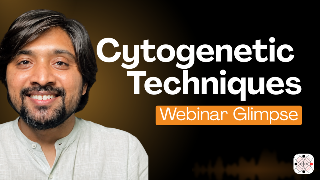
Resource Person: Dr. Tushar Chauhan, Ph.D., Founder & Director, Genetic Education
Date: 05 November 2026
Time: 11:00 AM – 1:00 PM
Platform: Genetic Education
Key Topics:
About the Webinar
The webinar “Cytogenetic Techniques: Karyotyping, FISH and Microarray Explained” was organized in association with the Department of Zoology, Gauhati University, and Genetic Education on 5 November 2026.
The session introduced undergraduate students and research scholars to the fundamentals of cytogenetics and its applications in clinical genetics. It focused on three important techniques used for chromosome analysis and the identification of genetic abnormalities: Conventional Karyotyping, Fluorescence In Situ Hybridization (FISH), and Chromosomal Microarray Analysis (CMA).
The objective was to provide students with an introductory understanding of how chromosomes are studied, how chromosomal abnormalities are identified, and how different cytogenetic techniques contribute to clinical diagnosis.
The session was delivered by Dr. Tushar Chauhan (Ph.D.), Founder and Director of Genetic Education, who has expertise in Karyotyping, FISH, Microarray, PCR, qPCR, DNA Sequencing, Next-Generation Sequencing (NGS), and Prenatal Genetics.
Introduction to Cytogenetics and Its Clinical Applications
The webinar began with an introduction to cytogenetics, a branch of genetics concerned with the study of chromosomes, their structure, number, and organization.
Students were introduced to the importance of chromosome analysis in understanding genetic conditions and identifying chromosomal abnormalities.
Cytogenetic investigations have important applications in clinical genetics, particularly in the diagnosis and investigation of:
- Numerical chromosomal abnormalities, such as trisomy and monosomy.
- Structural chromosomal abnormalities, including deletions, duplications, and translocations.
- Genetic conditions associated with chromosomal alterations.
- Prenatal and postnatal genetic abnormalities.
- Certain chromosomal changes associated with cancers and other clinical conditions.
The discussion highlighted how chromosome analysis connects fundamental genetics with clinical diagnosis and patient management.
Conventional Karyotyping: Understanding Chromosomes
The first major technique discussed was conventional karyotyping, one of the foundational methods used in cytogenetic analysis.
Karyotyping involves the examination of chromosomes under a microscope, generally using metaphase chromosome preparations. The chromosomes are arranged into homologous pairs based on their size, centromere position, and banding patterns.
A brief overview of the process included:
- Collection of an appropriate biological sample.
- Cell culture and stimulation, where required.
- Arresting dividing cells at metaphase.
- Hypotonic treatment and fixation.
- Preparation of chromosome slides.
- Chromosome staining and banding.
- Microscopic examination and chromosome arrangement.
Students were introduced to how a standard human karyotype contains 46 chromosomes arranged into 23 pairs, including 22 pairs of autosomes and one pair of sex chromosomes.
Advantages of Karyotyping
- Provides an overview of the complete chromosome complement.
- Helps identify numerical chromosomal abnormalities.
- Can detect large structural chromosomal alterations.
- Allows visualization of chromosome morphology and banding patterns.
Limitations of Karyotyping
- Requires dividing cells and suitable metaphase preparations.
- Has limited resolution compared with molecular cytogenetic techniques.
- Small deletions and duplications may remain undetected.
- Requires technical expertise in chromosome analysis and interpretation.
Fluorescence In Situ Hybridization (FISH)
The second technique introduced was Fluorescence In Situ Hybridization (FISH), a molecular cytogenetic technique that uses fluorescently labelled DNA probes to identify specific chromosome regions or DNA sequences.
Unlike conventional karyotyping, which provides a broader overview of chromosomes, FISH allows targeted investigation of particular genetic regions.
The basic principle involves hybridization of a fluorescently labelled DNA probe to its complementary DNA sequence in the sample. The resulting fluorescent signals are examined using a fluorescence microscope.
Key applications of FISH include:
- Detection of specific chromosomal deletions and duplications.
- Identification of selected chromosomal rearrangements.
- Detection of aneuploidy using chromosome-specific probes.
- Investigation of specific genetic abnormalities in prenatal and postnatal samples.
- Detection of selected chromosomal abnormalities in cancer genetics.
Advantages of FISH
- Provides targeted detection of specific genetic regions.
- Can be performed on interphase as well as metaphase cells.
- Offers better resolution than conventional karyotyping for selected targeted abnormalities.
- Can help investigate abnormalities that are difficult to characterize using routine chromosome banding.
Limitations of FISH
- Requires prior knowledge of the region or sequence being investigated.
- Detects only the abnormalities targeted by the selected probes.
- Does not provide a complete genome-wide assessment.
- Probe selection and signal interpretation require technical expertise.
Chromosomal Microarray Analysis (CMA)
The third technique discussed was Chromosomal Microarray Analysis, an important molecular cytogenetic approach used for genome-wide investigation of chromosomal copy number changes.
Unlike conventional karyotyping and targeted FISH, chromosomal microarray analysis evaluates DNA across numerous genomic regions simultaneously.
The technique uses thousands of DNA probes distributed across the genome to identify variations in DNA copy number.
Clinical applications of CMA include:
- Identification of submicroscopic deletions and duplications.
- Detection of copy number variations (CNVs).
- Investigation of individuals with developmental delay and intellectual disability.
- Evaluation of congenital anomalies.
- Genetic investigation in selected prenatal cases.
Advantages of CMA
- Provides genome-wide analysis of copy number variations.
- Detects smaller deletions and duplications that may not be visible through conventional karyotyping.
- Does not require dividing cells.
- Offers higher resolution for copy number changes compared with conventional chromosome analysis.
Limitations of CMA
- Generally does not detect balanced chromosomal rearrangements.
- Does not identify all types of genetic variations, such as most single-nucleotide variants.
- May identify variants of uncertain significance (VUS).
- Requires appropriate genetic interpretation and clinical correlation.
Comparison of Karyotyping, FISH and Microarray
An important part of the webinar was understanding how these three techniques differ and why different methods are selected for different clinical investigations.
| Feature | Karyotyping | FISH | Chromosomal Microarray |
|---|---|---|---|
| Basic principle | Microscopic chromosome analysis | Fluorescent DNA probe hybridization | Genome-wide DNA probe analysis |
| Analysis | Chromosome level | Targeted genomic regions | Genome-wide copy number changes |
| Sample requirement | Usually dividing cells | Interphase or metaphase cells | DNA from an appropriate sample |
| Numerical abnormalities | Yes | Yes, if targeted | Yes, through copy number analysis |
| Structural abnormalities | Large alterations | Specific targeted alterations | Deletions and duplications |
| Small deletions/duplications | Limited detection | Targeted detection | High-resolution detection |
| Balanced rearrangements | Generally detectable | Detectable with appropriate probes | Generally not detected |
| Main strength | Overall chromosome visualization | Targeted investigation | Genome-wide copy number analysis |
The comparison helped students understand that these techniques are not necessarily alternatives to one another. Each has specific strengths, applications, and limitations. The choice of technique depends on the suspected genetic abnormality and the clinical question being investigated.
Key Learning Outcomes
By the end of the webinar, participants were introduced to:
- The fundamentals of cytogenetics and its relevance to clinical genetics.
- The basic workflow of conventional chromosome preparation and karyotyping.
- The principles and applications of FISH.
- The role of chromosomal microarray in detecting copy number variations.
- The advantages and limitations of different cytogenetic techniques.
- The importance of selecting appropriate techniques for different genetic investigations.
Wrapping up:
Cytogenetic techniques continue to play an important role in clinical genetics, from conventional chromosome visualization to targeted molecular investigation and genome-wide copy number analysis.
Understanding the principles of Karyotyping, FISH, and Chromosomal Microarray Analysis provides students with a strong foundation for exploring modern cytogenetics and molecular diagnostics.
Through this collaborative academic session, Genetic Education and the Department of Zoology, Gauhati University, aimed to introduce students to the practical relevance of cytogenetic techniques and their applications in contemporary genetic research and clinical practice.
This webinar is a part of our free webinar series and the recording is available at Genetic Education Academy. Click the link to visit it: Webinar link
