
In some pregnancies, ultrasound findings raise concerns about the baby’s development, but chromosome analysis shows nothing unusual. So, what happens when the test says everything is normal, but something is still not right?
In this case, a 35-year-old pregnant woman was found to have several fetal abnormalities during the third trimester. The initial chromosome analysis appeared normal, but further investigation revealed a hidden chromosomal abnormality.
What was missed, and how was it finally identified?
Let’s explore this interesting prenatal case study.
Key Topics:
Case Background
Chromosomal deletions occur when a segment of a chromosome is lost. Depending on the size and location of the deletion, it may affect several genes and lead to developmental abnormalities, congenital defects, or intellectual disability.
Chromosome 11 contains many genes involved in normal growth and development. Deletions affecting its long arm, known as 11q deletions, can produce a wide range of clinical findings.
One well-known condition associated with a terminal deletion of chromosome 11q is Jacobsen syndrome. However, not all 11q deletions are terminal. Some involve an internal segment of the chromosome and are called interstitial deletions.
Interstitial 11q deletions are relatively uncommon, and their clinical effects can vary depending on the size and precise location of the missing segment.
Conventional G-banded karyotyping can detect many chromosomal abnormalities, but small deletions may be difficult to identify, especially when the chromosome bands appear otherwise normal. Array CGH provides a higher-resolution method for detecting gains and losses of DNA across the genome.
This case highlights the importance of combining conventional cytogenetics with molecular techniques when ultrasound findings suggest an underlying genetic abnormality.
Patient Summary
The case involved a 35-year-old woman who was pregnant for the first time. She and her partner were healthy and non-consanguineous, with no family history of congenital malformations or known genetic disorders.
The woman was referred for genetic counselling at 18 weeks of pregnancy.
Screening using cell-free DNA from maternal blood was negative for trisomy 13, trisomy 18, and trisomy 21.
However, a negative screening result for these common aneuploidies does not exclude all possible chromosomal abnormalities, including smaller deletions and duplications.
Prenatal Ultrasound Findings
At 28 weeks of pregnancy, a follow-up ultrasound revealed several abnormalities.
These included polyhydramnios, which means an increased amount of amniotic fluid, and intrauterine growth restriction, indicating that the fetus was smaller than expected for the gestational age.
The ultrasound also showed a persistent right umbilical vein and mild stenosis of the aortic arch, a narrowing involving part of the major artery that carries blood from the heart.
Together, these findings raised concerns about a possible underlying fetal abnormality.
The couple was offered further prenatal investigation to determine whether a chromosomal abnormality could explain the ultrasound findings.
Cytogenetic Investigation
Cordocentesis was performed at 28 weeks of gestation to obtain fetal blood for chromosome analysis.
G-banded Karyotyping
The fetal cells were cultured and examined using conventional G-banded karyotyping at approximately the 350-band level.
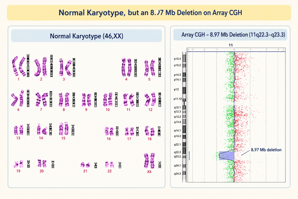
The result showed an apparently normal female karyotype: 46,XX
No obvious chromosomal abnormality was identified using this technique.
However, the ultrasound findings remained unexplained. The next step was to investigate the fetal DNA using a higher-resolution molecular cytogenetic technique.
Array CGH Findings
Array comparative genomic hybridization was performed using DNA extracted from uncultured fetal cord blood.
Unlike conventional karyotyping, which examines the appearance and banding pattern of chromosomes, array CGH compares DNA copy number across the genome to identify regions that have been gained or lost.
The array CGH result revealed a deletion of approximately 8.97 Mb on the long arm of chromosome 11, involving the region 11q22.3 to 11q23.3.
The result was reported as: arr[GRCh37] 11q22.3q23.3(107,686,511_116,660,613)x1
The notation x1 indicates that only one copy of this genomic region was detected instead of the usual two copies.
This finding established the presence of a submicroscopic deletion that had not been identified through conventional G-banded karyotyping.
Array CGH analysis of the parents did not identify the same deletion. The deletion was therefore classified as de novo, meaning it had arisen newly in the fetus rather than being detected as an inherited deletion in either parent.
No balanced chromosomal rearrangement or inversion was identified through parental G-banded karyotyping.
Genetic Explanation
Conventional G-banded karyotyping examines chromosomes under a microscope. It is useful for identifying changes in chromosome number and larger structural abnormalities. However, its ability to detect deletions depends on factors such as the size of the missing segment and the quality and resolution of the chromosome bands.
In this case, the deleted segment was approximately 8.97 Mb in size. Although this is a substantial region of DNA, the deletion was not recognized in the fetal karyotype at the resolution used.
Array CGH, on the other hand, evaluates DNA copy number across thousands of genomic locations. It can identify smaller losses and gains that may not produce an obvious change in the chromosome’s banding pattern.
The two methods therefore provide different types of information. Karyotyping shows the overall chromosome structure, while array CGH helps define the location and size of a DNA copy-number change.
The deletion in this case was interstitial, meaning that it involved an internal segment of chromosome 11 rather than extending to the end of the chromosome.
The deleted region contained approximately 30 genes with known or incompletely understood functions. However, because the pregnancy was terminated before birth and no autopsy was performed, it was not possible to establish a complete relationship between the deleted genes and the fetal findings.
Could Fragile Sites Have Played a Role?
The authors also discussed the possible role of chromosomal fragile sites in the formation of this deletion.
The deletion breakpoint was located near two known fragile regions on chromosome 11: FRA11B, a rare folate-sensitive fragile site, and FRA11G, a common fragile site that can be induced by aphidicolin.
Fragile sites are regions of chromosomes that can be particularly susceptible to breakage under certain conditions. Problems with DNA replication and chromosome stability may contribute to breaks in these regions.
Because the deletion was located near FRA11B and FRA11G, the authors proposed that regional chromosomal instability might have contributed to its formation.
However, this remains a proposed explanation. The study did not directly demonstrate that either fragile site caused the deletion.
Related article: An Introduction To Genome-Wide Association Study (GWAS)
Pregnancy Outcome
After genetic counselling, the couple decided to terminate the pregnancy, and termination was performed at 30 weeks of gestation.
A female fetus was delivered without apparent phenotypic abnormalities. However, an autopsy was not performed because the parents declined it.
As a result, the prenatal ultrasound findings could not be compared with a detailed postmortem examination, and the full clinical effects of the deletion remained uncertain.
This is an important limitation of the case. The genetic abnormality was clearly identified, but its complete relationship with the observed fetal abnormalities could not be established.
Key Learnings
This case provides several important lessons for clinical cytogenetics and prenatal diagnosis.
First, a normal conventional karyotype does not exclude every chromosomal abnormality. Smaller deletions and duplications may remain undetected, depending on their size and the resolution of the analysis.
Second, array CGH can identify genomic copy-number changes and define their approximate size and location. In this case, it detected an 8.97 Mb deletion involving chromosome 11q22.3 to 11q23.3.
Third, ultrasound abnormalities can provide an important reason to pursue further genetic testing even when routine screening or initial chromosome analysis is normal. Cell-free DNA screening for common aneuploidies does not rule out all other chromosomal abnormalities.
Finally, conventional karyotyping and array CGH should be viewed as complementary techniques. Karyotyping provides information about chromosome number and structure, while array CGH provides higher-resolution information about DNA gains and losses. Neither technique answers every genetic question, and the choice of test should be guided by the clinical findings.
Wrapping Up
This case demonstrates how an interstitial deletion of chromosome 11 can remain undetected by conventional G-banded karyotyping but be identified using array CGH.
The fetus had several abnormal ultrasound findings, including polyhydramnios, intrauterine growth restriction, a persistent right umbilical vein, and mild aortic arch stenosis. Although the karyotype was reported as 46,XX, array CGH revealed a de novo deletion of approximately 8.97 Mb at 11q22.3q23.3.
The case highlights the value of combining ultrasound assessment, conventional cytogenetics, and molecular cytogenetic testing to investigate suspected fetal chromosomal abnormalities.
For cytogenetics professionals, the key lesson is simple: when the clinical findings do not match the initial karyotype result, further molecular investigation is required.
Reference: Liu N, Yan J, Chen X, Song J, Wang B, Yao Y. Prenatal diagnosis of a de novo interstitial deletion of 11q (11q22.3 → q23.3) associated with abnormal ultrasound findings by array comparative genomic hybridization. Mol Cytogenet. 2014 Sep 25;7(1):62. doi: 10.1186/s13039-014-0062-y. PMID: 25298785; PMCID: PMC4189608.
>> Enroll in our free Cytogenetics for Beginners Course.