A 34-year-old woman with a twin pregnancy underwent genetic testing because it was considered a high-risk pregnancy.
The first-trimester NIPT reported a low risk for the common trisomies (trisomy 21, 18 and 13). However, the ultrasound evaluation showed some unusual findings.
One of the twins showed features suggesting a serious genetic condition, but the exact cause was not known at that stage. Further genetic investigations revealed an uncommon chromosomal abnormality, and the pregnancy was eventually terminated.
This interesting case gives us an opportunity to understand a rare structural chromosomal abnormality known as an isochromosome.
Key Topics:
Key Clinical Information
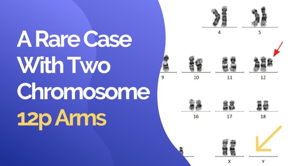
An isochromosome is a type of structural chromosomal abnormality in which a chromosome has two identical arms. It can have either two short arms (p arms) or two long arms (q arms).
Normally, a chromosome has one p arm and one q arm. In an isochromosome, one arm is lost and the other arm is duplicated. As a result, there is an imbalance in the genetic material.
For example, if an isochromosome contains two copies of the p arm, there will be an extra copy of genes located on that arm, while genetic material from the q arm may be missing.
Like many other chromosomal abnormalities, isochromosomes can occur randomly during cell division. Two commonly proposed mechanisms for their formation are centromere misdivision and U-type exchange.
Among these, centromere misdivision is one of the better-known mechanisms.
Normally, during cell division, chromosomes separate in a longitudinal direction, allowing the sister chromatids to separate correctly.
In the case of an isochromosome, the centromere may divide in the wrong direction, called transverse division. This can result in a chromosome carrying two copies of the same arm.
Isochromosomes have been reported in several chromosomal conditions. Some important examples include Pallister-Killian syndrome, which is associated with an isochromosome 12p, isochromosomes seen in some individuals with Turner syndrome, and certain hematological malignancies.
Patient Summary
A 34-year-old woman was carrying dichorionic diamniotic twins following IVF-ET.
There was no history of consanguineous marriage.
The first-trimester NIPT showed a low risk for trisomy 21, trisomy 18 and trisomy 13.
However, routine ultrasound detected an increased nuchal translucency of 3.4 mm in fetus A.
A detailed 3D ultrasound performed at 20 weeks showed additional abnormalities in fetus A, including:
- Limb shortening
- Mild lateral ventriculomegaly
Interestingly, fetus B appeared completely normal.
Because of these findings, further genetic investigations were recommended.
Genetic Investigations
Three different cytogenetic techniques were used in this case:
1. G-banding karyotyping
2. FISH (Fluorescence In Situ Hybridization)
3. Chromosomal microarray
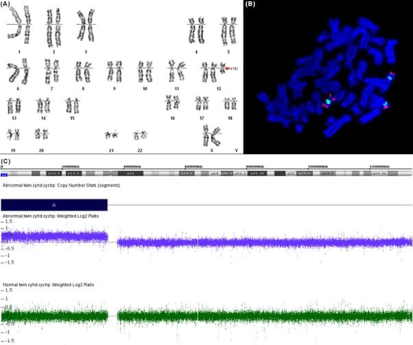
G-banding karyotyping was performed twice.
The first analysis was performed at 20 weeks using cultured amniocytes. A second analysis was performed at 23 weeks using fetal heart blood after termination of the pregnancy.
FISH was performed on the amniocytes using targeted probes. A red probe targeted 12p13.33, while a green probe targeted the centromeric region of chromosome 12.
A chromosomal microarray was also performed to investigate copy number changes across the genome.
It is important to note that NIPT had already been performed during the first trimester and had reported a low risk for the common trisomies.
The results from all these techniques provide an interesting comparison of what different genetic tests can reveal.
Key Outcomes
The first important finding was that the NIPT did not detect the abnormality.
G-banding karyotyping identified an isochromosome 12p in 80% of the analysed cells.
The reported karyotype was: 47,XX,i(12p)[40]/46,XX[10]
This indicates that 40 out of 50 analysed cells had the additional isochromosome 12p, while 10 cells had a normal female karyotype.
FISH supported the karyotyping findings and also detected the isochromosome in approximately 80% of cells.
Chromosomal microarray showed a copy number gain involving the 12p arm in approximately 80% of cells.
However, the result from the second karyotype performed after termination was different. The fetal heart blood showed approximately 34.3% mosaicism.
This difference is particularly interesting and leads us to an important concept in cytogenetics: tissue-specific mosaicism.
Genetics Explained
The present case represents an isochromosome 12p, which is associated with Pallister-Killian syndrome (PKS).
In this case, the fetus had an additional chromosome containing two copies of the 12p arm. This results in increased copy number of genes located on 12p, along with loss of the normal contribution from the corresponding 12q material on that abnormal chromosome.
The extra 12p material can affect several genes involved in development and may contribute to the clinical features associated with Pallister-Killian syndrome.
Previous studies have suggested that the isochromosome 12p may originate from a meiotic error and can be associated with maternal meiotic nondisjunction.
The copy number gain has been reported for the genes ING4, CHD4, ATN1, SOX9 and GATA6 located on the 12p arm. Notably, these genes play a pivotal role in development.
But there is another particularly interesting finding in this case.
The percentage of abnormal cells was not the same in different tissues. The amniocyte analysis showed around 80% abnormal cells, whereas the fetal heart blood showed only about 34.3% mosaicism.
This suggests that the isochromosome was distributed differently between tissues.
This is known as tissue-specific mosaicism, and it is an important feature of Pallister-Killian syndrome.
The level of mosaicism detected can therefore depend strongly on which tissue is tested.
Key Learnings
There are several important lessons from this case.
1. A negative NIPT does not rule out every chromosomal abnormality
NIPT is a powerful screening tool, particularly for common chromosome aneuploidies such as trisomy 21, 18 and 13. However, structural chromosomal abnormalities and some mosaic abnormalities may not be detected by NIPT.
This case is a good example of why an apparently low-risk NIPT result does not always explain abnormal ultrasound findings.
2. Different genetic techniques provide different information
- Karyotyping allowed the researchers to see and identify the abnormal chromosome.
- FISH provided targeted confirmation of the chromosome 12 abnormality.
- Chromosomal microarray helped identify the copy number gain involving 12p.
So, these techniques are not simply alternatives to each other. They can provide complementary information.
3. Mosaicism can be tissue-specific
This may be one of the most important lessons from this case.
The percentage of abnormal cells was different between amniocytes and fetal blood.
Therefore, the result obtained from one tissue may not necessarily represent the exact level of mosaicism in another tissue. This is particularly important when dealing with conditions such as Pallister-Killian syndrome.
4. Understanding chromosome structure matters
An isochromosome may look like a relatively simple structural abnormality, but it can create a complex copy number imbalance.
Understanding how the chromosome was formed helps us understand why certain genetic material is duplicated while other material is lost.
Wrapping Up
Isochromosome is a rare structural chromosomal abnormality that can lead to significant genetic imbalance and variable clinical features.
This case of isochromosome 12p is particularly interesting because it demonstrates three important concepts at the same time: Structural chromosome abnormality + mosaicism + tissue-specific variation.
It also reminds us that no single genetic test can answer every question. Thus, these three techniques are still important in cytogenetic investigations.
Reference:
Li, Lin et al. “Prenatal diagnosis of Pallister-Killian syndrome in one twin.” Clinical case reports vol. 6,8 1470-1473. 13 Jun. 2018, doi:10.1002/ccr3.1624.