
There is a rare chromosomal condition in which the patient has more than two complete sets of chromosomes. While most pregnancies affected by this condition do not survive, some babies are born alive but often die shortly after birth because of severe and complex abnormalities.
But this case is different.
An 18-month-old boy survived with four complete sets of chromosomes, giving him a total of 92 chromosomes in each cell. This is known as tetraploidy, and survival beyond infancy is extremely rare.
So, how did this happen, and how did this child survive for 18 months with such a rare chromosomal condition?
Let’s explore this unusual case of complete tetraploidy.
Read more: [Case study 10]: A Rare Case With Two Chromosome 12p Arms
Key Topics:
Key Clinical Information
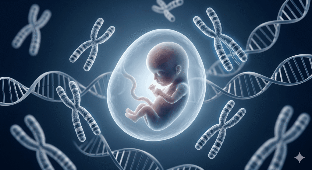
Tetraploidy is a rare chromosomal condition in which a cell contains four complete sets of chromosomes instead of the usual two. A normal human cell has 46 chromosomes, while a tetraploid cell has 92 chromosomes.
A complete tetraploid human karyotype can be written as 92,XXXX or 92,XXYY, depending on the sex chromosome composition.
Tetraploidy is usually not compatible with normal embryonic development. It is found in a small proportion of early spontaneous miscarriages, and most affected pregnancies end very early. Live-born children with complete tetraploidy are extremely rare, and most affected infants die shortly after birth or within the first year of life.
There are several possible mechanisms through which tetraploidy can occur.
One possible mechanism is a failure of cell division after DNA replication. During the very first fetal mitosis, DNA duplication happens as usual, but the cell cytoplasm does not divide into two daughter cells.
This traps all 92 chromosomes into a single cell, which further divides and forms the fetus.
Another possibility is the fertilization of an unreduced diploid egg by a normal sperm, or an unreduced diploid sperm fertilizing a normal egg. In this situation, the embryo starts with two complete chromosome sets from each parent and becomes tetraploid.
Tetraploidy can also occur through abnormal fertilization involving more than one sperm or through other early errors in chromosome duplication and cell division. These mechanisms are extremely rare and may lead to different forms of polyploidy depending on when the error occurs.

Patient Summary
This case describes an 18-month-old boy who was diagnosed with complete tetraploidy.
He was the second child of healthy, non-consanguineous parents. There was no significant family history of genetic or chromosomal disorders.
The pregnancy was initially considered normal, and prenatal ultrasound did not show any major structural abnormalities.
Prenatal and Birth History
The child was born at 40 weeks of pregnancy with a birth weight of 2,415 grams.
His Apgar scores were 5, 8, and 8.
Immediately after birth, he developed severe respiratory distress and required medical care.
Several congenital abnormalities and dysmorphic features were also observed. These included anophthalmia or microphthalmia, facial dysmorphism, and a 2 × 2 cm area of aplasia cutis on the scalp.
A loud heart murmur was also detected.
Craniofacial and Skeletal Findings
The child had several craniofacial and skeletal abnormalities.
He had a long cranium, a high forehead, hypoplastic and low-set ears, a long nose, and a small mouth.
Skeletal abnormalities included clubfoot and arachnodactyly, with unusually long fingers and long fingernails.
These findings, together with the other congenital abnormalities, suggested an underlying chromosomal disorder.
Neurological Findings
The neurological findings were severe.
The child had marked hypotonia, very low spontaneous activity, microcephaly, and profound psychomotor developmental delay.
He was also blind because of the severe abnormalities affecting the eyes.
Transfontanellar ultrasound showed small cysts in the lateral ventricles of the brain.
Overall, his neurological development was severely affected.
Cardiovascular Findings
Cardiac abnormalities were also present.
The child had a patent ductus arteriosus (PDA) and an atrial septal defect (ASD).
The heart was also unusually rotated.
These findings were part of the multiple congenital abnormalities associated with his tetraploid chromosome constitution.
Genital Findings
The external genitalia were hypoplastic.
He had a micropenis and small testes.
Together with the other clinical findings, this added to the overall picture of a severe chromosomal disorder.
Genetic Investigations
Because of the multiple congenital abnormalities and severe clinical presentation, cytogenetic testing was performed.
Peripheral blood lymphocytes were cultured and analyzed using conventional G-banded karyotyping at approximately the 550-band level.
The karyotype showed a complete tetraploid chromosome complement: 92,XXYY
Importantly, the tetraploid chromosome complement was not restricted to a small percentage of cells.
To confirm the finding, fibroblast culture was also performed. The fibroblast analysis confirmed complete tetraploidy without evidence of mosaicism.
This was therefore a case of complete, non-mosaic tetraploidy.
Build Skills- Advance Your Cytogenetics Career
Enroll in our structured Professional Clinical Cytogenetics Course and build practical skills in Karyotyping, FISH, Microarray and more.
Key Outcomes
Because of the severity of the clinical condition, treatment was mainly supportive and palliative.
The child received hospice care through the Warsaw Hospice for Children.
Further invasive cardiac and ophthalmic interventions were not performed because of the overall severity of his condition and poor prognosis.
Despite the severe abnormalities, he survived beyond 18 months and was cared for at home.
He continued to have severe feeding difficulties and required partial feeding through a tube or specialized teat. He also experienced recurrent respiratory infections.
Survival beyond the first year is unusual in complete tetraploidy, making this case particularly interesting from a clinical cytogenetics perspective.
Genetic Explanation
In a normal human embryo, each cell contains 46 chromosomes. During early development, DNA is replicated and the cell then divides so that each daughter cell receives the correct chromosome number.
In tetraploidy, this process goes wrong very early in development.
In this child, the most likely explanation is that the chromosomes were duplicated but the cell failed to divide properly. Instead of producing two cells with 46 chromosomes each, the cell retained all four chromosome sets, resulting in 92 chromosomes.
Because the abnormality occurred very early, the tetraploid chromosome complement was present throughout the tested tissues. This explains why both blood and fibroblast cultures showed complete tetraploidy rather than a mixture of normal and tetraploid cells.
The karyotype 92,XXYY means that the cells contained four complete chromosome sets, including two X chromosomes and two Y chromosomes.
But why is complete tetraploidy usually lethal?
The problem is not simply the increased number of chromosomes. Having four copies of the entire genome changes the dosage of thousands of genes at the same time.
During embryonic development, cells need very tightly controlled levels of gene expression. A complete extra set of chromosomes can disturb this balance and interfere with cell division, tissue development, organ formation, and normal growth.
This is why complete tetraploidy is usually incompatible with normal embryonic development and most affected pregnancies end in miscarriage.
However, this case is unusual because the child survived beyond 18 months despite having complete, non-mosaic tetraploidy.
Key Learnings
This case provides several important lessons about complete tetraploidy.
First, tetraploidy is a whole-genome chromosomal abnormality in which cells contain 92 chromosomes instead of 46. It usually occurs because of an early error in chromosome duplication or cell division.
Second, severe dysmorphic features, multiple congenital abnormalities, developmental delay, and unexplained multisystem involvement should raise suspicion of an underlying chromosomal abnormality.
Third, conventional G-banded karyotyping remains extremely important when polyploidy is suspected. A complete tetraploid genome can sometimes be missed or difficult to recognize using array CGH alone because array-based copy-number analysis compares DNA ratios across the genome. When both the test and reference samples have balanced whole-genome copy-number changes, the result may appear deceptively normal.
This means that a normal array result does not always exclude a whole-genome ploidy abnormality. The choice of genetic test is therefore important and should be guided by the clinical presentation.
Finally, this case shows that complete tetraploidy, although usually lethal early in development, can rarely be compatible with prolonged survival. The clinical outcome can therefore be more variable than expected in these extremely rare cases.
Read more: [Case Study #8] A Healthy Man With An Extra Chromosome
Wrapping Up
Complete tetraploidy is one of the rarest and most severe chromosomal abnormalities seen in humans. Most affected pregnancies end early, and live-born children usually have severe congenital abnormalities and a very poor prognosis.
This case is unusual because the child had complete, non-mosaic 92,XXYY tetraploidy and survived beyond 18 months.
The case also highlights the importance of conventional karyotyping in suspected chromosomal disorders. When a child presents with multiple congenital abnormalities and severe developmental problems, looking at the chromosome number directly can provide information that may not always be evident from molecular copy-number testing alone.
Reference: Bothur-Nowacka J, et al. Tetraploidy in the era of molecular karyotyping – What we need to remember. Pediatria Polska. 2013;88(5):467-471. doi:10.1016/j.pepo.2013.06.002.