A 7-year-old female patient was diagnosed with Turner syndrome.
Along with the classical clinical features of Turner syndrome (TS), she also exhibited several additional symptoms that suggested something unusual was happening at the chromosomal level.
Genetic investigations revealed overexpression of several X-chromosomal genes despite the absence of a normal second X chromosome.
This raises an interesting question.
If one X chromosome is missing, why are X-linked genes overexpressed?
Is the XIST gene dysfunctional?
And how can functional X chromosomal disomy occur in a patient with Turner syndrome?
Let’s explore an interesting case study that highlights an unusual combination of Turner syndrome, XIST dysfunction, and a small supernumerary marker chromosome (sSMC).
Key Topics:
Key Clinical Information
This case represents functional X chromosomal disomy in Turner syndrome caused by a small supernumerary marker chromosome (sSMC). In our previous case study, we discussed the basics of small supernumerary marker chromosomes and their clinical significance.
Normally, females have two X chromosomes, but one of them is transcriptionally silenced through a process known as X chromosome inactivation, mediated by the XIST (X-inactive specific transcript) gene.
This mechanism ensures that only one functional copy of the X chromosome remains active.
In this patient, the marker chromosome originated from the X chromosome but carried a functionally inactive or partially defective XIST region. As a result, the marker chromosome escaped complete X chromosome inactivation, allowing several X-linked genes to remain active.
This resulted in functional X chromosomal disomy, leading to overexpression of X-linked genes and additional clinical manifestations beyond the typical Turner syndrome phenotype.
Such cases are extremely rare and have been reported in only a small number of Turner syndrome patients.
Patient Summary
The patient was a 7-year-old girl diagnosed with Turner syndrome.
In addition to the characteristic Turner syndrome features, she presented with:
- Short stature
- Severe obesity
- Type 2 diabetes mellitus
Additional clinical findings included:
- Mild frontal bossing
- Broad nasal bridge
- Anteverted nostrils
- Low-set ears
- Full lips
- High-arched palate
- Severe nystagmus
- Divergent strabismus of the left eye
- Short neck
- Broad chest
- Widely spaced nipples
- Multiple nevi
- Delayed bone age
- Primary hypothyroidism
- Hypertriglyceridemia
- Hyperinsulinism
The patient also had intellectual disability, which is not typically considered a classical feature of Turner syndrome and therefore doctor prescribed further genetic evaluation.
Genetic Investigations
Several cytogenetic and molecular cytogenetic investigations were performed.
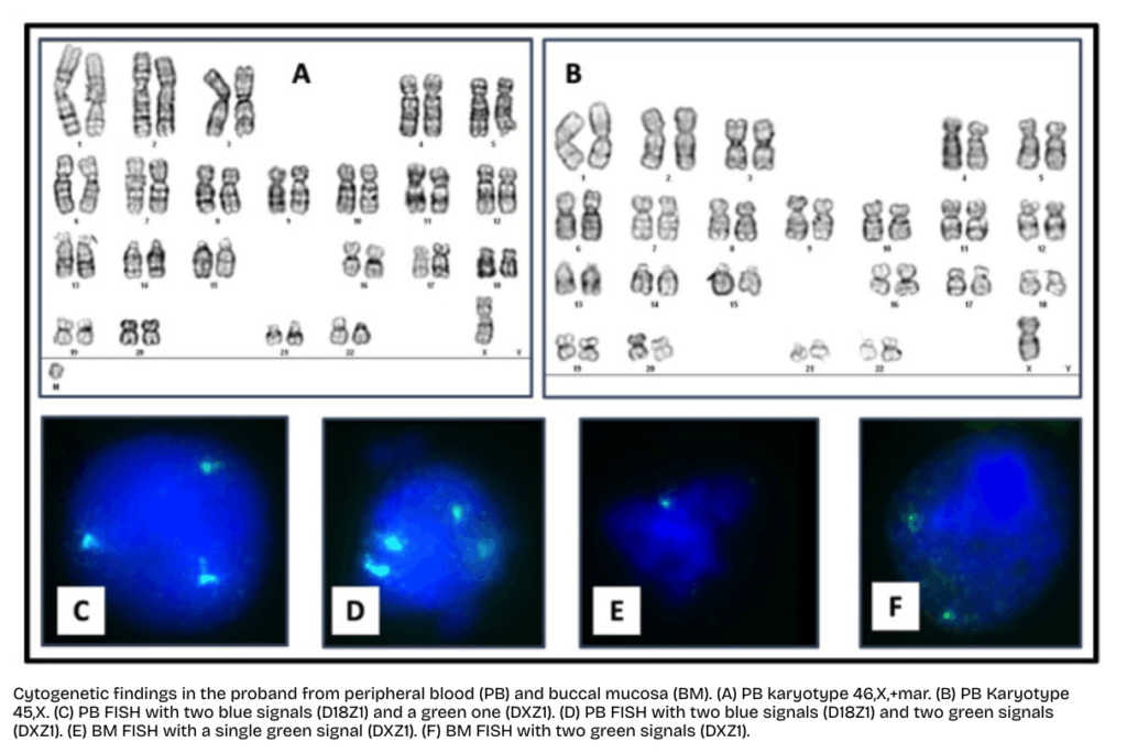
Conventional karyotyping with GTG banding was initially used to investigate the suspected Turner syndrome and revealed mosaicism along with a possible marker chromosome.
Fluorescence in situ hybridization (FISH) was performed on both peripheral blood and buccal mucosa samples to confirm the presence of the marker chromosome and evaluate tissue-specific mosaicism.
Array comparative genomic hybridization (array CGH) was subsequently performed to characterize the marker chromosome in greater detail, identify terminal deletions, and determine the genomic content of the sSMC.
Key Outcomes
Conventional karyotyping demonstrated mosaic Turner syndrome with two different cell populations: approximately 20% monosomy X and 80% cells containing an additional marker chromosome.
The reported karyotype was:
mos 46,X,+mar[16]/45,X[4]
Interphase FISH analysis further confirmed tissue-specific mosaicism.
In peripheral blood:
- 71.5% of nuclei showed a single X chromosome.
- 28.5% showed two X chromosome signals, indicating the presence of the marker chromosome.
However, the results differed in buccal mucosa.
In buccal epithelial cells:
- 32.6% of nuclei contained a single X chromosome.
- 67.4% demonstrated two X chromosome signals.
These findings clearly demonstrated tissue-specific mosaicism, indicating that the proportion of cells carrying the marker chromosome differed between tissues.
Array CGH further characterized the marker chromosome.
The sSMC measured approximately 25.34 Mb and showed terminal deletions at both ends of the X chromosome.
The identified deletions included:
Xp deletion: 56.85 Mb (Xpter–Xp11.21)
Xq deletion: 73.08 Mb (Xq21.1–Xqter)
These findings confirmed that the marker chromosome consisted primarily of the central region of the X chromosome with large terminal deletions.
Genetic Explained
To understand this case, two important concepts should be considered: X chromosome inactivation and small supernumerary marker chromosomes.
Normally, females possess two X chromosomes, but one of them becomes transcriptionally inactive through a process known as X chromosome inactivation. This process is controlled by the XIST gene, which produces a long non-coding RNA that coats the X chromosome and silences most of its genes.
In this patient, the marker chromosome originated from the X chromosome and retained the XIST locus. However, because of its abnormal structure, including extensive terminal deletions and possible ring chromosome formation, the XIST-mediated inactivation process was incomplete.
As a result, the XIST RNA was unable to fully coat and silence the marker chromosome. Consequently, several X-linked genes remained transcriptionally active, resulting in increased gene dosage despite the absence of a normal second X chromosome.
This phenomenon is known as functional X chromosomal disomy. Although the patient had Turner syndrome with only one normal X chromosome, the active marker chromosome behaved like an additional functional X chromosome, leading to overexpression of multiple X-linked genes.
This abnormal gene dosage likely contributed to the patient’s atypical phenotype, including intellectual disability, obesity, diabetes, and several dysmorphic features that are not commonly observed in classical Turner syndrome.
Key Learnings
This case highlights several important concepts in constitutional cytogenetics.
Conventional G-banding is often sufficient to diagnose Turner syndrome but is insufficient for complete characterization of marker chromosomes.
Molecular cytogenetic techniques such as FISH remain the gold standard for confirming the origin of an sSMC, while array CGH provides valuable information regarding genomic content and copy number variations.
In patients with Turner syndrome, the presence of additional findings such as intellectual disability, facial dysmorphism, metabolic abnormalities, or severe neurological features should immediately raise suspicion for a ring chromosome or an X-derived small supernumerary marker chromosome.
Another important observation from this case is tissue-specific mosaicism. The proportion of cells carrying the marker chromosome differed considerably between peripheral blood and buccal mucosa.
This demonstrates why multiple tissues should sometimes be evaluated in patients with mosaic chromosomal abnormalities.
Finally, this case illustrates the complementary role of karyotyping, FISH, and chromosomal microarray, each providing unique information required for establishing an accurate diagnosis.
Wrapping up
Small supernumerary marker chromosomes (sSMCs) are among the most complex constitutional chromosomal abnormalities. Their clinical presentation can range from completely normal individuals to patients with severe developmental disabilities.
The severity mainly depends on the chromosome of origin, the amount of euchromatin present, and the level of mosaicism.
This case also highlights that Turner syndrome patients with additional clinical features should always be investigated for a ring chromosome or an X-derived sSMC.
Finally, molecular cytogenetic techniques such as FISH and chromosomal microarray, along with conventional karyotyping, are essential for accurately diagnosing and characterizing such complex chromosomal abnormalities.
Reference:
González-Rodríguez, María Teresa Alejandra et al. “Identification of a Small Supernumerary Marker Chromosome in a Turner Syndrome Patient with Karyotype mos 46,X,+mar/45,X.” Genes vol. 14,2 253. 18 Jan. 2023, doi:10.3390/genes14020253.